Cardiac Imaging

While cardiac ultrasound is the widely used imaging modality for heart assessments, computed tomography (CT), magnetic resonance imaging (MRI) and nuclear imaging are also used and are often complimentary, each offering specific details about the heart other modalities cannot. For this reason the clinical question being asked often determines the imaging test that will be used.

Thumbnail

How cath labs can limit waste and become more sustainable

Making cardiac catheterization labs more efficient and less wasteful could help reduce the large carbon footprint associated with healthcare, according to a new commentary published in JACC: Cardiovascular Interventions.

Thumbnail

New U.S. guidelines on chronic coronary disease rethink use of beta-blockers, other medications

The updated recommendations from the American College of Cardiology and American Heart Association cover a variety of topics, including patient communication, follow-up imaging, nutritional supplements and more. 

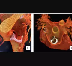
Image of a bioprosthetic valve within the RVOT from the SCCT's congenital heart disease guidelines

SCCT shares new recommendations on use of CT imaging to guide congenital heart disease treatment

The group collaborated with two other medical societies, SCAI and CHSS, on the expert consensus document. 

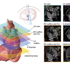
Axial planes suggested for screening the fetal heart at the time of the obstetric anatomic survey and as an initial series obtained during fetal echocardiography. #Fetalecho

ASE updates fetal echocardiography guidelines

The new 44-page document focuses on the detection, classification and risk assessment of congenital heart issues.

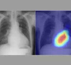
Artificial intelligence (AI) model using chest x-rays to evaluate cardiac function

No echocardiography, no problem? AI evaluates cardiac function using chest X-rays

Researchers think their new AI model could be especially helpful in areas where qualified physicians and/or ultrasound technology are in short supply.

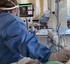
Video of American Society of Echocardiography (ASE) leadership explaining how the society recognized early the need for standardization and training for on and stepped forward to offer training for various point of care ultrasound (POCUS) users and to bring them into its membership. Image shows a critical care COVID patient being scanned with a Philips Lumify point-of-care ultrasound system with doctors on screen helping guide the user. #POCUS #ASE #ASE2023

ASE training clinicians as point-of-care ultrasound grows

The American Society of Echocardiography recognized the lack of training and standardization for point-of-care ultrasound users and decided to do something about it. 

Thumbnail

Tracking patient outcomes after valve-in-valve TAVR, redo SAVR

It is becoming more and more important, researchers noted, to examine the lifetime management of patients who undergo aortic valve replacement.

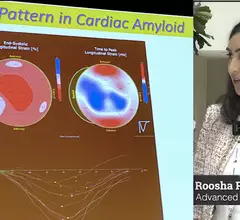
Roosha Parikh, MD, advanced imaging cardiologist, St. Francis Heart Hospital, Long Island, New York, and a clinical assistant professor of medicine at the State University of New York at Stony Brook, presented in one of the ASE 2023 amyloid sessions and spoke with Cardiovascular Business about the disease.

Amyloidosis now a hot topic in cardiac imaging due to new drug treatment

Advanced cardiac imager Roosha Parikh, MD, explained that cardiac amyloidosis is regularly misdiagnosed. It can often be identified in echocardiography results, however, if physicians know how to identify it. 

Around the web

Ron Blankstein, MD, professor of radiology, Harvard Medical School, explains the use of artificial intelligence to detect heart disease in non-cardiac CT exams.

Eleven medical societies have signed on to a consensus statement aimed at standardizing imaging for suspected cardiovascular infections.