The routine use of intravascular imaging (IVI) for guiding percutaneous coronary intervention (PCI) procedures is highly recommended, according to a new guidance from the American College of Cardiology (ACC). These technologies provide clinicians with additional information that cannot be adequately captured by coronary angiography alone.
The new analysis, written by the ACC’s Interventional Council, was published in full in the Journal of the American College of Cardiology.[1] It describes all currently available IVI modalities, reviews the latest clinical research and calls for all cardiac cath labs (CCLs) to embrace this technology going forward.
“Over the last several decades, there have been significant reductions in catheter size and improvements in both imaging quality and catheter deliverability,” wrote first author Alexander G. Truesdell, MD, an interventional cardiologist with Virginia Heart and the Inova Heart and Vascular Institute, and colleagues. “However, despite multiple randomized clinical trials, meta-analyses, and registries that support the use of IVI to guide PCI, the overall adoption of IVI remains low, with current estimates demonstrating usage in <15% of all PCI procedures with significant operator and center variation. Various explanations have been proposed for the underutilization of IVI, including unfamiliarity with imaging equipment, knowledge gaps in image interpretation, perception of increased procedural times adversely affecting workflow efficiency, lack of adequate financial reimbursement, and an evidence base in need of further definitive clinical trials.”
What are the primary intravascular imaging modalities?
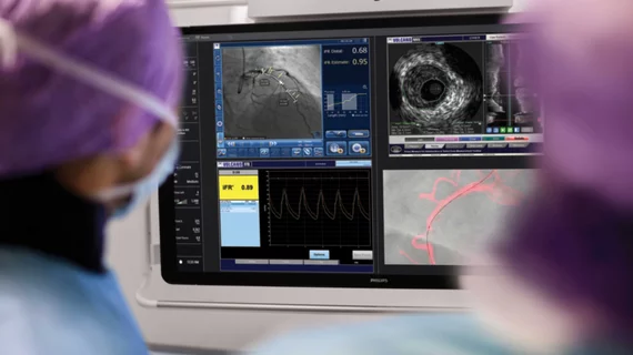
The ACC guidance highlights three IVI modalities: intravascular ultrasound (IVUS), optical coherence tomography (OCT) and near-infrared spectroscopy (NIRS).
IVUS involves the use of intravascular catheters that generate ultrasound pulses, providing real-time images for the clinician to review. IVUS technology has improved over time, the authors noted, leading to improved image resolution.
OCT catheters emit and receive near-infrared light waves, which are converted into real-time 3D images. Its resolution is the best of any IVI modality by a significant margin, but it does require the target vessel to be cleared of blood with a saline or contrast flush. It also lacks the tissue depth penetration of IVUS. More recently, vendors have started developing hybrid IVUS-OCT systems capable of delivering both IVUS and OCT with a single catheter.
The authors describe NIRS as “a unique IVI modality” based on near-infrared light being absorbed and scattered. NIRS systems use an IVUS-sized catheter and use electromagnetic radiation to evaluate tissues based on light absorption in the near-infrared spectrum. A spectrographic analysis of the returning light from the vessel wall can tell the cardiologist the composition of the plaques
IVUS vs. OCT
Clinicians are regularly faced with a key decision: IVUS or OCT? According to the ACC’s recommendations, each option has its benefits. IVUS does not require the target vessel to be cleared of blood, for example, making it a better fit for performing chronic total occlusion revascularization or treating chronic kidney disease. IVUS is also sometimes preferred when imaging larger vascular structures due to its deeper tissue imaging capability.
OCT, on the other hand, can deliver high-resolution imaging if the vessel lumen, plaque, remodeling and the stent struts better than IVUS. It is also a better fit for measuring and characterizing heavily calcified stenoses, the authors noted.
The many uses of IVI
IVI can help interventional cardiologists image a patient’s vessels prior to PCI, guide stent deployment and expansion during PCI and confirm the procedure was a success following PCI.
“IVUS and OCT improve the detection, localization, quantification and characterization of coronary calcification (thickness, angle and length) to guide adjunctive therapies, such as angioplasty, atherectomy and lithotripsy, to ensure sufficient calcium fracture to facilitate stent delivery and adequate stent expansion,” the authors wrote. “IVUS and OCT-based scoring systems have been devised to identify calcified stenoses at risk for stent underexpansion (a cause of stent thrombosis and restenosis) that may necessitate adjunctive calcium modification before stent implantation.”
The group also noted that IVI “may help optimize the overall number and length of stents deployed and shorten procedure times.” In addition, it has been found that IVI helps “significantly reduce” the use of contrast materials during PCI procedures.
Another key benefit of IVI modalities is that they can help clinicians study new medications and medical devices.
Key IVI recommendations from the ACC
The ACC’s interventional experts recommend that IVI be treated as “an essential adjunct to angiography for specific lesion subsets or any scenario where angiography may inadequately elucidate anatomy.” In addition, they wrote, IVUS and OCT should be available in all CCLs throughout the United States.
“Initial and ongoing training and education” are also highly recommended for all operators to ensure they know how to interpret IVI images whenever necessary. On a similar note, the authors called for IVI to be prioritized by training programs and professional societies throughout the United States.
“The use of IVI has become an important and essential adjunct to the practice of conventional PCI, but further innovation and optimizations are necessary to realize the full potential of IVI during interventional procedures,” the authors concluded.
This is just a tip of the iceberg when it comes to the contents of this thorough analysis from the ACC. Read the full document here.

Michael has more than 16 years of experience as a professional writer and editor. He has written at length about cardiology, radiology, artificial intelligence and other key healthcare topics.