Philips premieres new 3D MRI technique, aims to bring speed and efficiency to cardiac imaging
Royal Philips and the Spanish National Center for Cardiovascular Research (CNIC) have developed a new cardiac MRI protocol they say can provide a full evaluation of the heart in just minutes.
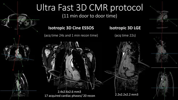
The updated technique, known as Enhanced SENSE by Static Outer-volume Subtraction (ESSOS), was designed so that it can be used with existing equipment. According to Philips, users can now acquire full, accurate—the 3D images of the heart much faster than with any previous protocols.
“In just over 20 seconds, all the information needed to know the shape and function of the heart has been acquired,” Javier Sánchez-González, PhD, a CNIC member who also works for Philips, said in a prepared statement. “And if you need to evaluate the degree of fibrosis after cardiac muscle death, another 20-second acquisition is all it takes, completing the cardiac study in less than a minute.”
The company also sees this as a way to improve efficiency and prioritize patient comfort. Cardiologist Sandra Gómez-Talavera, MD, a CNIC researcher, added that MRI scans are still not as common in cardiology as other modalities. Her team aims to help change that trend with this ongoing collaboration.
“The main cause is the time needed to do a full study,” she said. “A complete study requires about an hour, a period that causes many patients not to finish the test due to the discomfort it causes them.”
Though this new protocol is still being finalized, the parties hope that it can soon be implemented in clinical settings throughout the world.
Gómez-Talavera, Sánchez-González and colleagues wrote about this new protocol at length in JACC: Cardiovascular Imaging. Click here to read their full analysis.
Additional information is available on the Philips website.

