Echocardiography

Cardiac ultrasound uses reflected sound waves (echos) to create images of anatomy inside the body. Echocardiograms are the primary cardiac imaging modality used to assess the heart and diagnose or track cardiac issues. Echo is the gold standard imaging modality to assess the heart, particularly with calculating left ventricular ejection fraction (LVEF), which is a measure of cardiac output. In addition to noninvasive standard transthoracic echo (TTE), invasive transesophgeal echo (TEE) is also used when clearer, more detailed imaging of the heart is needed. Both 3D and 4D echo echo systems are rapidly gaining wider adoption and enable new types of assessments, especially in the structural heart space and in transcatheter procedural guidance. Find news on general ultrasound imaging.
FDA has now cleared more than 1,000 AI models, including many in cardiology

Cardiology is the medical speciality with the second most FDA clearances overall. 

James N. Kirkpatrick, MD, a veteran cardiologist and cardiac imaging specialist with the University of Washington, died unexpectedly on Jan. 1. He was 54 years old.

Cardiologist remembered for his ‘great empathy, love and generosity’

James N. Kirkpatrick, MD, a cardiologist with the University of Washington, died unexpectedly on Jan. 1. Colleagues have shared loving tributes to Kirkpatrick, highlighting his passion for ethics and strong leadership skills.

Thumbnail

New AI program delivers rapid, accurate echo video assessments

The new technology shows early potential to make a significant impact on imaging workflows and patient care. 

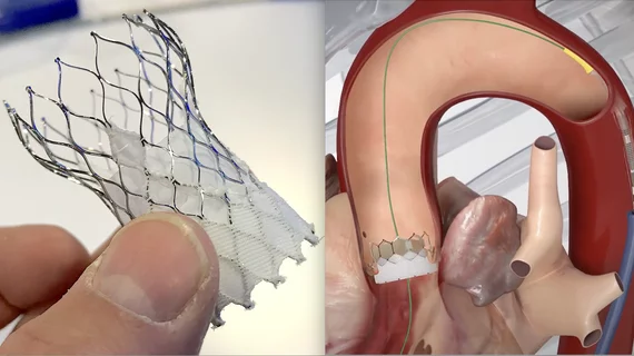
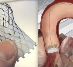
Medtronic's Evolut Pro TAVR valve treating aortic stenosis

Medtronic TAVR valves at heart of key studies presented during PCR London Valves 2024

One study presented during the three-day conference focused on the potential benefits of an optimized TAVR pathway. Another study, meanwhile, tracked changes in paravalvular leak severity over time.

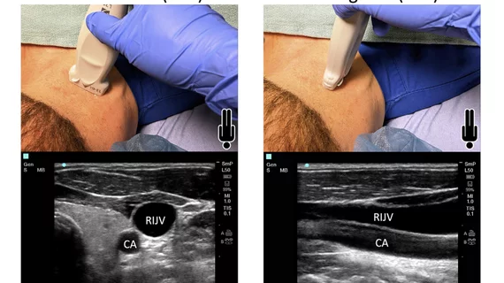
Gilbert H. Tang, MD, professor in the Department of Cardiovascular Surgery at the Icahn School of Medicine and surgical director of the Structural Heart Program at the Mount Sinai Health System, explains how to overcome the limited imaging windows of the tricupid valve using a TEE and intracardiac echo (ICE) at TCT 2024.

How to overcome imaging challenges during tricuspid valve procedures

Gilbert Tang, MD, surgical director of the structural heart program at the Mount Sinai Health System, discussed a key challenge care teams often encounter during tricuspid valve procedures. 

The rapid rise of artificial intelligence (AI) has helped cardiologists, radiologists, nurses and other healthcare providers embrace precision medicine in a way that ensures more heart patients are receiving personalized care.

FDA clears AI screening tool for cardiac amyloidosis

Ultromics designed EchoGo Amyloidosis to evaluate routine echocardiogram results for signs of cardiac amyloidosis. It received the FDA's breakthrough device designation back in 2023.

The iCardio.ai EchoMeasure AI software to automate measurements for cardiac echo imaging. It was FDA cleared in October 2024.

FDA approves AI startup’s new software for echo assessments

iCardio.ai, co-founded by a cardiologist, is part of the Cedars-Sinai Accelerator program. The startup has already announced new partnerships with Viz.ai, Abbott and others in the cardiac health space. 

Ultrafast/high frame rate myocardial contrast echocardiography

Ultrafast myocardial contrast echocardiography shows early potential to evaluate CAD

Ultrafast MCE could go on to become a go-to treatment option for obstructive coronary artery disease, according to the authors of a new first-in-human clinical study.

Around the web

Several key trends were evident at the Radiological Society of North America 2024 meeting, including new CT and MR technology and evolving adoption of artificial intelligence.

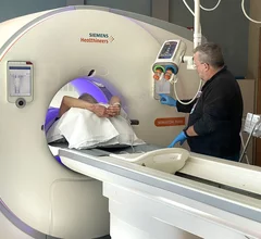
Ron Blankstein, MD, professor of radiology, Harvard Medical School, explains the use of artificial intelligence to detect heart disease in non-cardiac CT exams.