Echocardiography

Cardiac ultrasound uses reflected sound waves (echos) to create images of anatomy inside the body. Echocardiograms are the primary cardiac imaging modality used to assess the heart and diagnose or track cardiac issues. Echo is the gold standard imaging modality to assess the heart, particularly with calculating left ventricular ejection fraction (LVEF), which is a measure of cardiac output. In addition to noninvasive standard transthoracic echo (TTE), invasive transesophgeal echo (TEE) is also used when clearer, more detailed imaging of the heart is needed. Both 3D and 4D echo echo systems are rapidly gaining wider adoption and enable new types of assessments, especially in the structural heart space and in transcatheter procedural guidance. Find news on general ultrasound imaging.
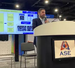
Jeremy Slivnick, MD, presents at the American Society of Echocardiography (ASE) 2023 meeting on how artificial intelligence (AI) can help make echocardiography better able to detect subtle signs of early cardiac amyloid disease when it is easier to treat with better outcomes. ssistant professor of medicine and an advanced cardiac imager at the University of Chicago.

AI models for cardiac amyloidosis could make a world of difference

Jeremy Slivnick, MD, spoke with Cardiovascular Business about AI's potential to transform how cardiac amyloidosis is diagnosed and treated. 

Tootsie Roll paravalvular leak transcatheter heart valves

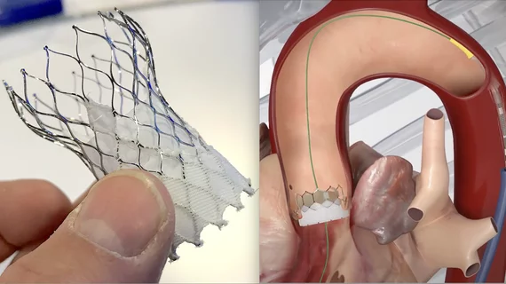
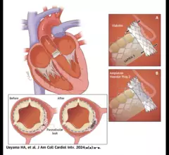
New ‘Tootsie Roll’ technique could help cardiologists treat PVL in transcatheter heart valves

Paravalvular leak remains a significant problem after TAVR, SAVR and even TMVR. Could a new-look interventional technique help heart teams overcome this issue and improve patient outcomes? 

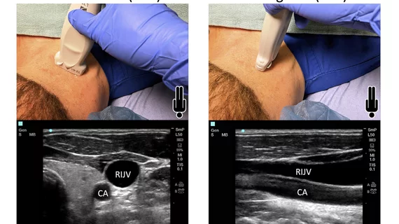
ICE-guided Watchman procedures linked to safety concerns—is TEE better for LAAO or will things improve?

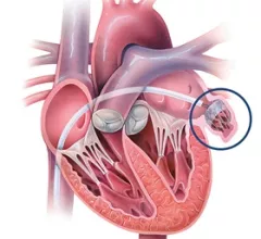
TEE and ICE are each associated with certain advantages and disadvantages, researchers explained. The new analysis included data from nearly 40,000 patients.

Craig Sable Children's National Hospital AI echo

AI spots signs of rheumatic heart disease in echocardiograms as well as cardiologists

Researchers think this represents a potential breakthrough for low- and middle-income countries where access to high-quality imaging evaluations is still limited. 

Self-expandable TAVR cusp-overlap ICE

ICE guidance reduces risk of permanent pacemaker implantation after TAVR with a self-expandable valve

Heart teams can limit the risk of conduction disturbances that lead to permanent pacemaker implantation by utilizing both the cusp-overlap method and intracardiac echocardiography.

artificial intelligence consultation

Imaging specialists partner with Pfizer to deliver AI-powered cardiac amyloidosis evaluations

Improving care for patients with cardiac amyloidosis has emerged as one of the hottest topics in cardiology. Pfizer helped kickstart that trend in 2019 when it gained FDA approval for two separate medications for the rare, but potentially fatal disease. 

TAVR after mitral valve replacement

TAVR after mitral valve replacement linked to positive outcomes, but heart teams must plan ahead

Researchers in India performed TAVR on two relatively young patients with a preexisting mechanical mitral valve, sharing their experience in a new case report. The group highlighted the importance of planning ahead.

transesophageal echocardiography (TEE)

New expert guidelines: Start PHV evaluations with echocardiography, but other imaging modalities can provide value

New guidelines from the American Society of Echocardiography, made in collaboration with the Society for Cardiovascular Magnetic Resonance and Society of Cardiovascular Computed Tomography, represent an update of the group's original recommendations from 2009.

Around the web

Several key trends were evident at the Radiological Society of North America 2024 meeting, including new CT and MR technology and evolving adoption of artificial intelligence.

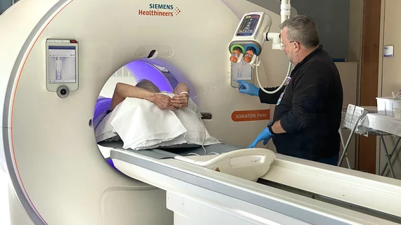
Ron Blankstein, MD, professor of radiology, Harvard Medical School, explains the use of artificial intelligence to detect heart disease in non-cardiac CT exams.