Echocardiography

Cardiac ultrasound uses reflected sound waves (echos) to create images of anatomy inside the body. Echocardiograms are the primary cardiac imaging modality used to assess the heart and diagnose or track cardiac issues. Echo is the gold standard imaging modality to assess the heart, particularly with calculating left ventricular ejection fraction (LVEF), which is a measure of cardiac output. In addition to noninvasive standard transthoracic echo (TTE), invasive transesophgeal echo (TEE) is also used when clearer, more detailed imaging of the heart is needed. Both 3D and 4D echo echo systems are rapidly gaining wider adoption and enable new types of assessments, especially in the structural heart space and in transcatheter procedural guidance. Find news on general ultrasound imaging.
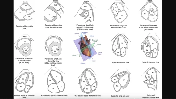
Axial planes suggested for screening the fetal heart at the time of the obstetric anatomic survey and as an initial series obtained during fetal echocardiography. #Fetalecho

ASE updates fetal echocardiography guidelines

The new 44-page document focuses on the detection, classification and risk assessment of congenital heart issues.

Artificial intelligence (AI) model using chest x-rays to evaluate cardiac function

No echocardiography, no problem? AI evaluates cardiac function using chest X-rays

Researchers think their new AI model could be especially helpful in areas where qualified physicians and/or ultrasound technology are in short supply.

Video of American Society of Echocardiography (ASE) leadership explaining how the society recognized early the need for standardization and training for on and stepped forward to offer training for various point of care ultrasound (POCUS) users and to bring them into its membership. Image shows a critical care COVID patient being scanned with a Philips Lumify point-of-care ultrasound system with doctors on screen helping guide the user. #POCUS #ASE #ASE2023

ASE training clinicians as point-of-care ultrasound grows

The American Society of Echocardiography recognized the lack of training and standardization for point-of-care ultrasound users and decided to do something about it. 

Roosha Parikh, MD, advanced imaging cardiologist, St. Francis Heart Hospital, Long Island, New York, and a clinical assistant professor of medicine at the State University of New York at Stony Brook, presented in one of the ASE 2023 amyloid sessions and spoke with Cardiovascular Business about the disease.

Amyloidosis now a hot topic in cardiac imaging due to new drug treatment

Advanced cardiac imager Roosha Parikh, MD, explained that cardiac amyloidosis is regularly misdiagnosed. It can often be identified in echocardiography results, however, if physicians know how to identify it. 

Stephen Little, MD, discusses trends in echocardiography at ASE 2023. #ASE23 #ASE2023 What is new in cardiac ultrasound.

Back in the spotlight: Exploring echocardiography's revival

American Society of Echocardiography President Stephen Little, MD, says several trends and technologies are coming together at once, leading to renewed interest in echo.

PHOTO GALLERY: The trends and technologies at ASE 2023

Browse through some of the many highlights from the American Society of Echocardiography's 2023 annual meeting. 

Roberto Lang, MD, explains how AI can help novice users get diagnostic quality cardiac echo exams at ASE 2023.#ASE #AI #ASE2023

AI can help novice sonographers deliver optimal echocardiograms

Roberto Lang, MD, has been closely involved with the development of AI technology that helps inexperienced ultrasound users perform high-quality echo exams. "It is an unbelievable thing when you see it working," he said. 

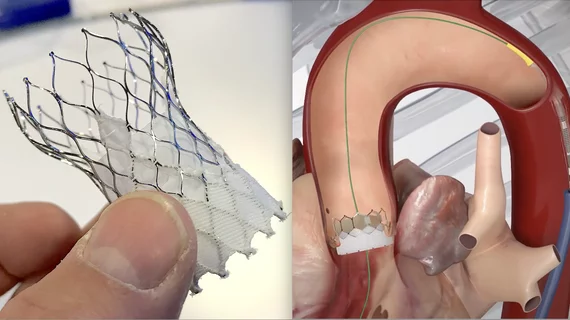
Balloon expansion deployment of an Edwards Lifesciences Sapien 3 transcatheter aortic valve replacement (TAVR) device.

TAVR vs. surgery: Exploring 5-year outcomes among intermediate-risk patients

A group of renowned TAVR specialists examined data from 783 pairs of intermediate-risk patients, sharing their findings in the Journal of the American College of Cardiology.

Around the web

Ron Blankstein, MD, professor of radiology, Harvard Medical School, explains the use of artificial intelligence to detect heart disease in non-cardiac CT exams.

Eleven medical societies have signed on to a consensus statement aimed at standardizing imaging for suspected cardiovascular infections.

Trimed Popup
Trimed Popup