Echocardiography

Cardiac ultrasound uses reflected sound waves (echos) to create images of anatomy inside the body. Echocardiograms are the primary cardiac imaging modality used to assess the heart and diagnose or track cardiac issues. Echo is the gold standard imaging modality to assess the heart, particularly with calculating left ventricular ejection fraction (LVEF), which is a measure of cardiac output. In addition to noninvasive standard transthoracic echo (TTE), invasive transesophgeal echo (TEE) is also used when clearer, more detailed imaging of the heart is needed. Both 3D and 4D echo echo systems are rapidly gaining wider adoption and enable new types of assessments, especially in the structural heart space and in transcatheter procedural guidance. Find news on general ultrasound imaging.
Zio AT mobile cardiac telemetry device

FDA warning letter: iRhythm committed multiple violations, failed to report patient deaths in time

“It is your firm’s responsibility to ensure compliance with applicable laws and regulations administered by FDA,” according to the warning letter. These issues, the agency added, “may be symptomatic of serious problems in your firm’s manufacturing and quality management systems.”

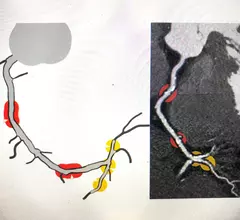
An example of HeartFlow's new RoadMap Stenosis software that uses artificial intelligence (AI) to show areas of interest for possible stenting based on a patient's CT scan and FFR-CT. This software is still undergoing beta testing at several hospitals and will likely be rolled out commercially later in 2023.

Cardiology has embraced AI more than most other specialties

Cardiology is linked to the second largest group of FDA-cleared clinical AI algorithms, and the number is still growing. 

artificial intelligence AI deep learning

AI model predicts LVEF during routine coronary angiograms

The video-based deep neural network showed potential for limiting invasive exams and improving patient care. 

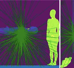
X-ray photon trajectory during the simulation phantom study from the side and top views. Due to scattering of the X-rays when they hit the lower end of the patient bed, exposure in mainly to the lower body of the interventional echocardiographer performing transesophageal echocardiography. The green lines are the scattered photon trajectories calculated by Monte Carlo simulation in the study.

Radiation exposure in the cath lab: Tracking the impact on interventional echocardiographers

Researchers found that echocardiographers in the cath lab are exposed to high doses of radiation on the right half of their body, especially the waist and lower body. 

New ASE guideline outlines training standards for interventional echocardiography

The American Society of Echocardiography (ASE) released a new guideline document that outlines uniform training standards for interventional sonographers guiding structural heart procedures.

artificial intelligence AI deep learning

Cardiologists find AI to be more accurate than sonographers at interpreting echocardiograms

Overall, cardiologists made corrections to 16.8% of AI-generated assessments and 27.2% of sonographer assessments.

Echocardiography expert Patricia A. Pellikka, MD, discussed the trend of increasing artificial intelligence (AI) integration in cardiac ultrasound with Cardiovascular Business at American College of Cardiology (ACC) 2023 meeting.

AI's growing impact on echocardiography

Cardiology has the second largest number of FDA-cleared AI algorithms, and many of them are for cardiac ultrasound. Echocardiography expert Patricia A. Pellikka, MD, discusses this trend and how AI is helping improve echo.

A figure from the ASE pediatric POCUS guidelines showing parasternal short-axis view demonstrating RV dilatation with bowing of the septum into the left ventricle, indicating pulmonary hypertension in this child with shock and pertussis.

New ASE guidance examines cardiac POCUS in children

The recommendations were developed by a writing group comprised of experts from different specialties, offering an array of perspectives and approaches.

Around the web

Ron Blankstein, MD, professor of radiology, Harvard Medical School, explains the use of artificial intelligence to detect heart disease in non-cardiac CT exams.

Eleven medical societies have signed on to a consensus statement aimed at standardizing imaging for suspected cardiovascular infections.

Trimed Popup
Trimed Popup