New ‘Tootsie Roll’ technique could help cardiologists treat PVL in transcatheter heart valves
Paravalvular leak (PVL) is a common issue after transcatheter aortic valve replacement (TAVR), surgical aortic valve replacement (SAVR) and transcatheter mitral valve replacement (TMVR), but cardiologists have struggled to find an effective treatment strategy for those patients.
With that ongoing challenge in mind, researchers have developed a new “Tootsie Roll” technique specifically designed to address PVL related to transcatheter heart valves (THVs). The group presenting its initial findings in JACC: Cardiovascular Interventions.[1]
“The presence of PVL can lead to disabling symptoms of heart failure and hemolysis, and mild or greater PVL after TAVR is associated with an increased risk of long-term mortality,” wrote senior author Vasilis Babaliaros, MD, an interventional cardiologist with the Emory Structural Heart and Valve Center in Atlanta, and colleagues. “Transcatheter PVL closure has emerged as an attractive treatment option because surgical PVL closure involves redo sternotomy and high perioperative mortality, ranging from 6% to 10%. Several registries have evaluated transcatheter PVL closure using various available devices and have shown promising results. However, PVL related to THVs is underrepresented in these registries.”
A new technique for treating transcatheter heart valve-related paravalvular leak
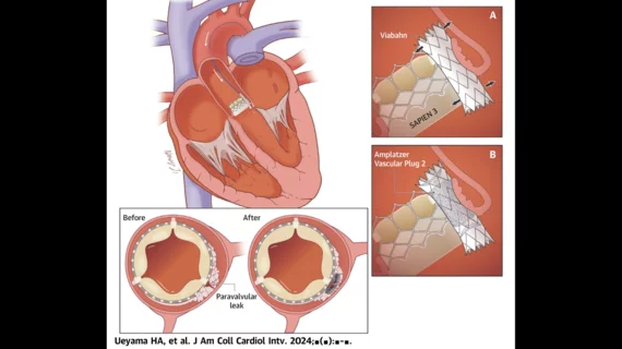
The group’s Tootsie Roll technique involves covering the PVL with a nonpermeable covered stent—they used a Viabahn device from Gore—and then “transforming the complicated PVL tract into a cylinder” so that an Amplatzer vascular plug from Abbott could be deployed, promoting in-stent hemostasis.
“Targets with a tunneled, tortuous tract, or crescent-shaped PVLs were considered unsuitable for closure with available devices and were expected to benefit most from this procedure,” the authors wrote. “We observed that these characteristics are frequently found in PVLs associated with THVs.”
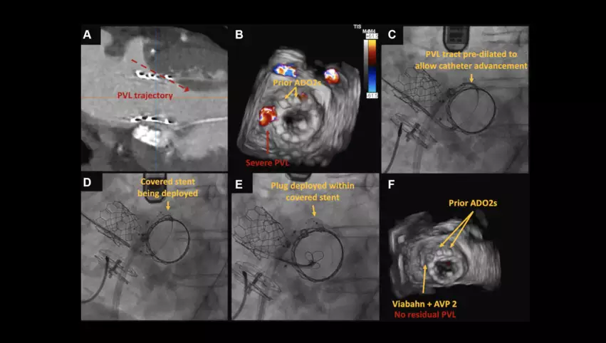
Both transesophageal echocardiography (TEE) and contrast-enhanced CT scans were used to help heart teams plan ahead of the procedures, ensuring the stents were the right diameter and length. The stent’s diameter was oversized on purpose, the group explained, “so that it would deform itself to cover the entire contour of the PVL, especially in the case of a crescent-shaped PVL.” Guidance during each procedure was provided by TEE and fluoroscopy, and operators also relied on a guide catheter, an angled guidewire, a vertebral catheter and a guidewire to advance the covered stent to its destination. After a shuttle sheath was advanced through the stent and into the patient’s left ventricle, the vascular plug was finally deployed within the stent. Echocardiography and angiography helped confirm PVL closure was a success.
Treating mitral PVL with the Tootsie Roll technique in which a covered stent is inserted into the area of the leak to turn the hole into a cylinder shape that is easier to treat. An Amplatzer vascular plug is then used to occlude the covered stent and seal the PVL. Images courtesy of Ueyama et al.tootsie roll technique paravalvular leak
Early outcomes with the Tootsie Roll technique
Babaliaros et al. tracked data from eight patients treated with this new technique at a single facility. All procedures were a technical success, all patients were discharged alive and there was no residual PVL except for in one patient with mild regurgitation “due to another untreated PVL location.”
One TMVR patient who was asymptomatic at the time of discharge did die within 30 days. There were no rehospitalizations due to heart failure, and no patients showed signs of hemolysis. After seven months, the team added, the seven surviving patients showed no or just trace signs of PVL.
“We demonstrated that the Tootsie Roll technique: 1) is technically feasible with 100% success rate in various THVs in both the aortic and mitral position; 2) is safe with no in-hospital complications or postprocedure hemolysis; and 3) provides a significant reduction of PVL to less than mild in all cases,” the authors wrote.
Key points to remember about the Tootsie Roll technique
The group emphasized a few points at the end of their analysis. For example, they explained, this technique is only recommended for patients with PVL tracts that can be covered with a single covered stent. In addition, there is a risk of stent migration during or after deployment; “appropriate sizing” of the covered stent is crucial to keep that risk to a minimum.
The group also noted that this procedure will be associated with higher costs due to the use of both a covered stent and a vascular plug.
“The long-term cost-effectiveness, which involves potential for reintervention in cases of significant residual PVL, remains uncertain until larger-scale, longer-term follow-up studies are conducted,” the authors wrote.
Finally, because they were only exploring initial results of this new procedure, the research team highlighted the fact that much more research with many more patients is still necessary.
Click here to read the full study.


