FDA has now cleared more than 1,000 AI models, including many in cardiology
The U.S. Food and Drug Administration (FDA) has now cleared more than 1,000 clinical artificial intelligence (AI) algorithms to be used commercially for direct patient care in the United States. Cardiology is No. 2 among all healthcare specialties with 161 FDA clearances; some of those are even approved for multiple specialties.
Radiology is by far the king of AI FDA clearances with 758 algorithms, making up about 76% of all clinical AI in the U.S. Neurology comes in at an extremely distant third place with 35 algorithms. There are 15 other specialities with cleared AI, but they each number less than 20 algorithms.
The FDA updated its AI-enabled device approval list in late December, which showed the agency technically reached the 1,000 mark back in September. The first AI algorithm was cleared in 1996, and the number of submissions to the FDA has accelerated very rapidly in the past few years. The agency is now clearing an average of about 20 AI algorithms per month, and the FDA says that number is expected to rise in the coming years.
The FDA's latest update includes clearances made in the period July 30 to Sept. 27, 2024. Of the 42 new approvals over that two-month period, 10 were specific to cardiology:
• EchoGo Heart Failure (2.0) from Ultromics Limited. It is an automated diagnostic aid for patients undergoing routine echo assessment to help detect heart failure with preserved ejection fraction (HFpEF).
• HeartKey Rhythm from B-Secur Limited. It analyses single lead ECG data from adults on compatible devices for arrhythmia diagnostics, including Holters, extended Holters, ECG patches and event recorders. It has an application program interface (API) to provide ECG signal conditioning, ECG signal quality assessment, QRS beat detection, ventricular ectopic beat detection and ECG rhythm analysis.
• Acumen Assisted Fluid Management (AFM) software feature from Edwards Lifesciences. The software provides physiological insight into a patient's estimated response to fluid therapy and the
associated hemodynamics. It is intended for use in surgical patients that require advanced hemodynamic monitoring. Fluid administration suggestions are offered to the clinician.
• CAC (gated) Algorithm from BunkerHill Health. This AI offers opportunistic calcium scoring assessments to be performed automatically on patients undergoing any type of chest CT that includes the heart in the field of view. It detects the presence and estimates the quantity of coronary artery calcium (CAC) in ECG gated, non-contrast CT scans.
• Fetoly-Heart from Diagnoly. The AI analyzes fetal ultrasound image sequences to automatically detect heart views and quality criteria within the views. It is indicated for use during routine fetal heart examination of 2nd and 3rd trimester pregnancy (gestational ages from 17 to 40 weeks).
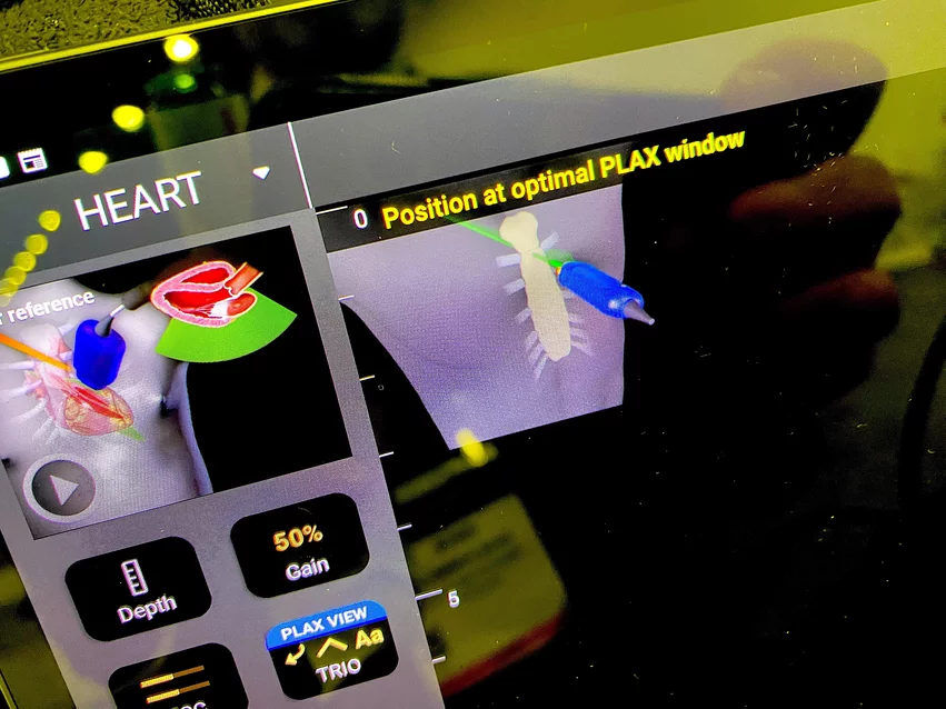
• AI Platform 2.0 (AIP002) from Exo Imaging. AI automates ideal image detection, measurements and calculations for structures and functions during echo and lung examsIt also offers an image quality feedback score for the sonographer.
• Kosmos from EchoNous, Inc. AI automates the acquisition, processing, measurements of cardiac ultrasound synchronized with ECG and digital auscultation sounds. Includes anatomical labeling and automated left ventricular ejection fraction.
• HealthCCSng from Nanox AI Ltd. Automated, opportunistic AI screening that detects coronary artery calcification on any non-gated CT scans of the chest that include the entire heart.
• AISAP Cardio V1.0 from Aisap. Automatically processes and analyzes acquired cardiac point of care ultrasound (POCUS) images, producing a report with diagnostic assessment and measurements of several key cardiac structural and functional parameters This includes presence of valvular regurgitation and stenosis, and left ventricle ejection fraction (LVEF).
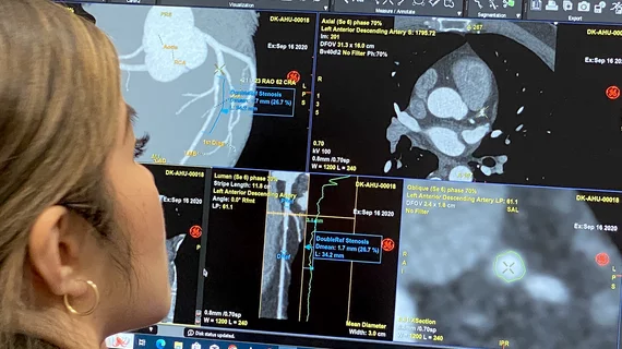
• CardIQ Suite from GE Healthcare. AI offers automated cardiovascular anatomy and pathology assessments on 2D or 3D CT cardiac non-contrast and angiography. It provides capabilities for the visualization and measurement of vessels and visualization of chamber mobility to aids in diagnosis and treatment paths for coronary artery disease, functional parameters of the heart, heart structures and follow-up for stent placement, bypasses and plaque imaging. It provides calcium scoring, which can be used with non-contrasted cardiac images, to evaluate calcified plaques in the coronary arteries, heart valves and great vessels.


