3D-printed sugary stent boosts speed of suturing arteries
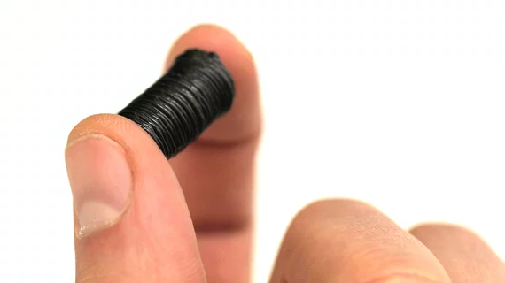
A team of researchers has developed a sugar-based stent that provides structural support during challenging microvascular suturing surgeries. The stent, which is 3D printed to match the diameters of specific arteries, fits inside the adjacent ends of a blood vessel while the stitching process takes place—and then quickly dissolves after blood flow resumes.
Researchers led by University of Nebraska engineer Ali Tamayol, PhD, designed the stent and published their early experience with the device in October in the journal Advanced Healthcare Materials.
Stent-assisted suturing of ex vivo pig arteries took about five minutes, significantly trimming the roughly 15 minutes it normally takes to perform microanastomosis with a conventional clamp-based technique, according to a University of Nebraska press release. The researchers said they eventually plan to test the stent in live animal arteries, with the long-term goal of 3D printing the device at hospitals to match the dimensions of individual patients.
According to the release, the team used a glucose derivative called dextran to provide the right balance of strength and flexibility. Sucrose and sodium nitrate were also added to help prevent blood clotting.
“One thing that I really like about this concept: We are always trying to avoid sugar,” Tamayol said. “Everyone knows that sugar is (sometimes) bad. But here we found an application in which it’s good.”

