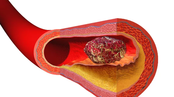
New technique for battling blood clots uses nanodroplets, ultrasound imaging
Researchers have developed a new strategy for treating especially challenging blood clots, sharing their findings in Microsystems & Nanoengineering.
The technique, still under evaluation before clinical testing can begin, involves using nanodroplets and a tiny “ultrasound drill” to break down retracted blood clots from the inside out. When liquid perfluorocarbons inside the nanodroplets convert into gas, they become microscopic bubbles; those bubbles then disrupt the clot, causing it to dissolve.
“The use of ultrasound to disrupt blood clots has been studied for years, including several substantial studies in patients in Europe, with limited success,” co-author Paul A. Dayton, PhD, a professor of biomedical engineering at both the University of North Carolina at Chapel Hill and North Carolina State University, said in a statement. “However, the addition of the low-boiling point nanodroplets, combined with the ultrasound drill has demonstrated a substantial advance in this technology.”
Overall, the group’s in vitro tests indicate that this strategy could outperform previous methods for dissolving blood clots—but there is still a lot of work to do before anything can be tested on human patients in a clinical setting.
Looking ahead, the research team plans to begin pre-clinical testing with animal models as they evaluate their strategy’s potential use as a treatment for deep vein thrombosis.
A grant from the National Institutes of Health funded the team’s work. Read their full analysis in Microsystems & Nanoengineering here.

