Hybrid imaging predicts adverse events for patients with CAD
Hybrid imaging with coronary CT angiography (CCTA) and single photon emission CT (SPECT) can provide powerful prognostic value for patients being evaluated for coronary artery disease, according to a single-center study published online July 3 in Radiology.
Researchers from University Hospital Zurich followed 375 patients for a median of 6.8 years after they had undergone imaging evaluation for CAD. All patients were assessed for stenosis (at least 50 percent upon CCTA) and ischemia upon SPECT myocardial perfusion imaging at baseline.
The annual rate of death or myocardial infarction was 7 percent for patients with evidence of both stenosis and ischemia, compared to 3.7 percent for those meeting one of these criteria and 1.2 percent for those who met neither. Annual rates of major adverse cardiac events—a composite of death, MI, unstable angina requiring hospitalization and coronary revascularization—were 21.8 percent, 9 percent and 2.4 percent for these groups, respectively.
“Throughout the full follow-up of 10 years, the results have consistently identified patients with a matched finding at hybrid imaging to have the worst outcome, while a normal finding confers a favorable long-term prognosis, confirming the excellent risk stratification ability of cardiac hybrid imaging in patients who are known to have or suspected of having CAD,” wrote senior study author Philipp A. Kaufmann, MD, director of cardiac imaging at University Hospital Zurich, and colleagues.
The researchers said an “unmatched” finding—in which one of the imaging modalities was consistent with CAD—still is associated with a threefold higher risk of death or MI and may warrant more aggressive risk factor modification in those patients.
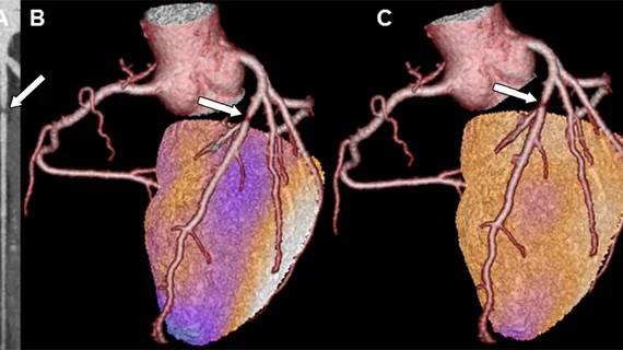
The SPECT and CCTA images were fused together for analysis, allowing researchers to simultaneously assess patients’ degree of stenosis and severity of stress-induced ischemia.
According to Kaufmann, the results suggest a CCTA could be used for an initial evaluation of patients with suspected CAD. If the findings are normal, no additional imaging is necessary. But if a significant lesion is present, nuclear imaging may be helpful to assess ischemia, make a hybrid image and more accurately quantify risk.
"The strategy of direct referral to invasive coronary angiography without noninvasive imaging is obsolete," Kaufmann said in a press release. "Even after documenting coronary artery disease with coronary CT angiography, we need further noninvasive evaluation before deciding upon revascularization versus medication."

