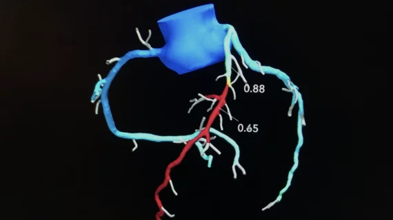
CT-FFR before TAVR improves detection of coronary artery disease, limits invasive imaging exams
Performing CT-derived fractional flow reserve (CT-FFR) before transcatheter aortic valve replacement (TAVR) improves the accuracy of coronary CT angiography (CCTA) and helps limit unneeded invasive coronary angiography (ICA), according to a new study published in JACC: Cardiovascular Interventions.[1]
Screening TAVR patients for coronary artery disease (CAD) before their procedure is crucial due to the negative impact CAD can have on clinical outcomes, the study’s authors explained, but such screening typically includes ICA.
“ICA is indicated for the pre-TAVR assessment of CAD,” wrote first author Joyce Peper, MSc, with the department of radiology at St. Antonius Hospital and department of cardiology at University Medical Center Utrecht, and colleagues. “If abnormal or equivocal, fractional flow reserve (FFR) could be used to complement ICA. Alternatively, noninvasive evaluation of the coronary arteries, which are visualized in the routine computed tomography procedure for the assessment of the access route and for sizing of the valve prosthesis, has been suggested for the detection of significant CAD. CCTA has good sensitivity (95%), moderate specificity (65%), and an acceptable diagnostic accuracy to rule out hemodynamically significant CAD in a preprocedural screening.”
CT-FFR is a new imaging method that is noninvasive and does not lead to any additional radiation exposure In fact, CT-FFR was described as a “reasonable” and “effective” treatment option for diagnosing CAD in the American Heart Association and American College of Cardiology’s 2021 chest pain guidelines.
“CT-FFR is based on the principles of computational fluid dynamics to simulate invasive FFR, the reference diagnostic test for the diagnosis of CAD,” the authors explained. “There is no need for additional testing because CT-FFR can be added to CCTA without changes in imaging protocols, radiation dose, or the addition of medication. Also, the use of noninvasive diagnostic tools prevents complications associated with ICA, such as stroke, vascular events, bleeding, myocardial infarction, and major thrombosis.”
Prior studies had found that CT-FFR can improve the accuracy of CCTA, so Peper et al. wanted to see if this association could improve care for TAVR patients. The group examined data from more than 300 patients with severe symptomatic aortic valve stenosis who underwent TAVR work-up from 2015 to 2019. Each patient underwent CCTA and ICA within three months.
Overall, the sensitivity, specificity, positive predictive value, negative predictive value and diagnostic accuracy per patient were 76.9%, 64.5%, 34%, 92.1%, and 66.9%, respectively, for CCTA alone and 84.6%, 88.3%, 63.2%, 96%, and 87.6%, respectively, for a CT-FFR-guided approach.
Also, the authors calculated that ICA could have been avoided in 57.1% of patients with a CT-FFR-guided approach; this is significantly more than the 43.6% of patients associated with a CCTA-guided approach.
“This study supports the use of CCTA and CT-FFR as gatekeepers for ICA work-up for TAVR patients,” the authors wrote. “Furthermore, evidence is provided for the additional value of CT-FFR over CCTA diameter stenosis only.”
The team emphasized that additional research is still needed to confirm their findings. Also, it is unclear what a history of percutaneous coronary intervention or coronary artery bypass graft may mean for this relationship.
Related CT-FFR and TAVR Content:
Imaging update: CT-FFR is safe and feasible for patients with severe aortic stenosis
VIDEO: HeartFlow FFR-CT sees increased interest after inclusion in the 2021 Chest Pain Guidelines
Late balloon valvuloplasty safe and effective for patients with THV dysfunction
Transcaval TAVR outperforms transaxillary TAVR when femoral access is not an option
TAVR outcomes unaffected when women require a smaller valve prosthesis
Reference:

