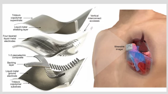
New stamp-sized wearable device uses AI to deliver on-the-go cardiac imaging
A new wearable ultrasound device developed by engineers and physicians out of the University of California San Diego (UCSD) is the size of postage stamp, but it could end up making a massive impact on patient care.
“The increasing risk of heart diseases calls for more advanced and inclusive monitoring procedures,” corresponding author Sheng Xu, a professor of nanoengineering at UCSD, said in a prepared statement. “By providing patients and doctors with more thorough details, continuous and real-time cardiac image monitoring is poised to fundamentally optimize and reshape the paradigm of cardiac diagnoses.”
The device, designed to be worn for up to 24 hours at a time, generates real-time ultrasound images of the heart and uses artificial intelligence (AI) to process data on how much blood the heart is pumping. It is made of stretchable material and adheres directly to a person’s skin. According to the team of specialists at UCSD, the device can even be worn when the user is exercising, providing an accurate look at the heart’s function during a time when complications are more likely to stand out.
Xu et al. also noted that their device provides high-quality imaging results without exposing the patient to any radiation, which can’t be said about other modalities such as CT.
Additional details on the wearable cardiac ultrasound
Co-author Ruixiang Qi, a master’s student at UCSD, provided some context about the device’s AI capabilities in the same prepared statement.
“The imaging-segmentation deep learning model is the first to be functionalized in wearable ultrasound devices,” he said. “It enables the device to provide accurate and continuous waveforms of key cardiac indices in different physical states, including static and after exercise, which has never been achieved before.”
Also, co-author Hongjie Hu, a researcher at UCSD, emphasized just how crucial it is for researchers to keep improving the cardiac imaging technology available to clinicians.
“The heart undergoes all kinds of different pathologies,” Hu said. “Cardiac imaging will disclose the true story underneath. Whether it be that a strong but normal contraction of heart chambers leads to the fluctuation of volumes, or that a cardiac morphological problem has occurred as an emergency, real-time image monitoring on the heart tells the whole picture in vivid detail and visual effect.”
The group’s full analysis is available in Nature.[1]

