Radiologists select 2023's top images in cardiothoracic imaging
Radiology: Cardiothoracic Imaging, part of the Radiological Society of North America (RSNA) family of journals, has shared its list of the Top 2023 Images in Cardiothoracic Imaging.[1] A team of the group’s trainee editors selected the winners.
“The 2023 winner and runners-up reflect the vibrant and dynamic nature of cardiothoracic imaging, as well as the role of state-of-the-art imaging in the diagnosis of clinically important, rare, and potentially life-threatening cardiothoracic and vascular conditions,” wrote first author Domenico Mastrodicasa, MD, a radiologist and cardiovascular imager with the University of Washington, and colleagues. “Cardiothoracic imaging continues to evolve, fueled by technical innovations, such as dynamic chest radiography, digital tomosynthesis, and dark-field radiography.”
Images were selected based on whether or not they involved the use of a new technology, were educational or were visually compelling. Manuscripts published from July 2022 to June 2023 were eligible.
The winning cardiothoracic images
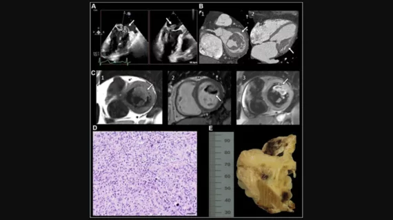
The winning images were a part of the article “Undifferentiated Cardiac Sarcoma of the Mitral Valve: Multimodal Imaging Assessment,” which appeared in Radiology: Cardiothoracic Imaging in April.[2]
The article focused on a 75-year-old female who underwent transesophageal echocardiography (TEE). Radiologists identified a “lobulated intracardiac mass” on the patient’s mitral valve, and a full multimodality imaging assessment was performed. The mass was confirmed to be a high-grade undifferentiated pleomorphic sarcoma.
“The article exemplifies the multimodality noninvasive imaging assessment of cardiac masses, using transesophageal echocardiography for initial diagnosis and to evaluate the impact of the mass on valve function, cardiac CT for submillimeter isotropic spatial visualization, and cardiac MRI for tissue characterization,” the authors wrote. “This approach allowed a comprehensive insight into different aspects of this rare and visually striking mass.”
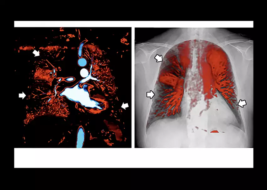
Two images from "Dynamic Chest Radiography of Acute Pulmonary Thromboembolism" by Yamasaki et al. (Left) Multiple segmental perfusion defects (white arrows) are shown in the iodine map created from dual-energy imaging captured using dual-layer spectral CT. (Right) Well-defined triangular perfusion defects (white arrows) are visible in the bilateral lungs in the perfusion image of dynamic chest radiography, similar to the iodine map. Image/caption courtesy of RSNA.
Other top images in cardiothoracic imaging from 2023
The article "Dynamic Chest Radiography of Acute Pulmonary Thromboembolism,” published in August 2022, was selected as the competition’s first runner-up due to its fascinating images.[3] The study’s authors examined how perfusion imaging can be used to evaluate a 50-year-old female presenting with acute pulmonary embolism.
“Key highlights of this article include the novelty of the technology and the visual appeal of pulmonary perfusion defects demonstrated with multiple imaging modalities,” the authors wrote.
These represent just some of the images highlighted by Mastrodicasa et al. Click here for the full breakdown.


