
AI creates accurate 4D heart scans in seconds
Researchers have developed and validated a new artificial intelligence (AI) model capable of producing four-chamber cardiac MR (CMR) images in seconds, publishing their findings in European Radiology Experimental.[1]
A team of cardiology and imaging experts with the University of East Anglia (UEA) led the research, with assistance from specialists with the University of Sheffield, University of Leeds, Norfolk and Norwich University Hospitals NHS Foundation Trust and Leiden University Medical Center. The group already has extensive experience working in the world of 4D MRI technology.
For this latest analysis, lead researcher Dr. Pankaj Garg, an associate professor of cardiovascular medicine at UEA, and colleagues developed a fully automated deep learning algorithm for time-resolved segmentation of CMR images. It was trained on data from more than 800 patients and then validated with data from another 101 patients. The mean age of the validation cohort was 54 years old, and 65% were male.
Overall, the group found that its new-look AI model delivered four-chamber evaluations comparable to the work of trained radiologists with “minimal bias.”
“The AI model precisely determined the size and function of the heart's chambers and demonstrated outcomes comparable to those acquired by doctors manually but much quicker,” Garg said in a prepared statement. “Unlike a standard manual MRI analysis, which can take up to 45 minutes or more, the new AI model takes just a few seconds. This automated technique could offer speedy and dependable evaluations of heart health, with the potential to enhance patient care.”
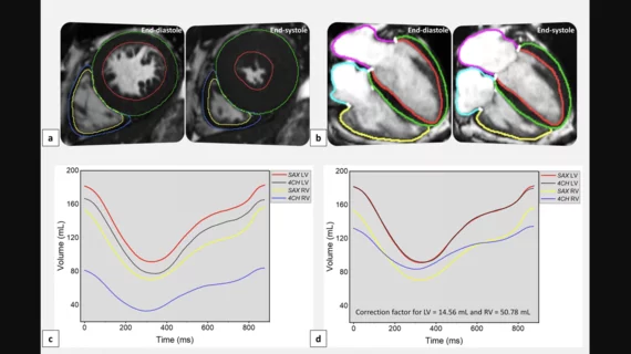
Garg et al. noted that they took specific steps to achieve its low levels of bias. For instance, an initial version of their AI model underestimated both left and right ventricular volumes compared to a ground-truth short-axis cine analysis. By applying two correction factors, that “systematic bias” was minimized, leading to much more effective CMR evaluations.
Click here to read the full study.

Michael has more than 18 years of experience as a professional writer and editor. He has written at length about cardiology, radiology, artificial intelligence and other key healthcare topics.