
CT-Guided PCI: Extending the Value of CCTA into the Cath Lab
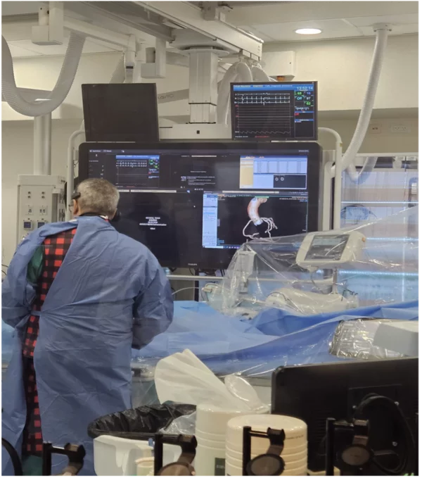
Traditionally, the interventional cardiologist first visualizes the coronary anatomy in detail when contrast is injected in the catheterization laboratory. For many patients, however, that anatomy has already been captured, often in considerable detail, by coronary CT angiography (CCTA) performed earlier in the clinical pathway. This article considers how this dataset can be used to plan the procedure, an approach referred to as CT-guided PCI.
This situation is becoming increasingly common. The 2021 AHA/ACC multisociety chest pain guideline gives CCTA a Class 1 recommendation for intermediate- to high-risk patients with stable chest pain and no known coronary artery disease. CCTA is also incorporated into the evaluation of selected intermediate-risk patients with acute chest pain without high-risk features [1]. European guidance is concordant: the 2024 ESC guidelines for chronic coronary syndromes recommend CCTA as the preferred test to rule out obstructive coronary artery disease in patients with a low to moderate pretest likelihood [2]. These recommendations concern diagnosis rather than procedural planning, but they mean that a CCTA dataset is now available for a growing proportion of patients referred for PCI.
This raises a practical question: If detailed three-dimensional coronary anatomy is already available, how can it be used to plan PCI more effectively?
Starting with the right view
Invasive coronary angiography remains fundamental to PCI, but it depicts three-dimensional anatomy as a series of two-dimensional projections. Vessel overlap and foreshortening can obscure lesion length, bifurcations and side branches, and the true spatial relationship between adjacent coronary segments.
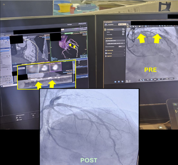
CCTA provides the coronary geometry before the procedure begins. The coronary tree can be evaluated from multiple orientations to identify projections that minimize foreshortening and vessel overlap. This enables one of the most intuitive applications of CT-guided PCI: selection of an appropriate C-arm angulation before the first angiographic acquisition, rather than through successive angiographic runs [3,4].
In a prospective multicenter registry of 102 patients with suspected single-vessel CAD, CCTA helped correctly identify the non-obstructive coronary artery in all patients. A CCTA-guided ultraselective ICA strategy was estimated to reduce procedure time by 49%, radiation exposure by 35%, and contrast volume by 33% [5].
In another single-center, open-label randomized pilot trial of 30 patients undergoing PCI for aorto-ostial RCA lesions, CT-derived C-arm angles were used for procedural planning in the intervention arm. Optimal stent positioning was achieved in all CT-planned patients, whereas geographic mismatch occurred in five of 15 patients treated with conventional angiographic guidance. Contrast volume, radiation exposure and procedure time were also lower in the CT-planned group [6].
Planning the intervention before crossing the lesion
Once the vessel has been characterized in three dimensions, the value of CT extends beyond angulation.
CCTA can provide information on lesion length, vessel dimensions and tapering, plaque distribution, and potential proximal and distal landing zones. These data may be particularly relevant when angiographic assessment is limited by foreshortening or diffuse disease. In ostial lesions, a precise understanding of the vessel origin and lesion extent may help avoid geographic miss; in long lesions, CT can show where disease begins and ends before stent length is selected [3,4].
An early randomized single-center trial from 2011, found that adding preprocedural CCTA planning influenced PCI strategy, including longer stented segments, and improved lesion coverage by intravascular ultrasound criteria [7].
The same principle applies to bifurcation disease. Three-dimensional visualization can clarify the bifurcation angle and the course of the main vessel and side branch, the distribution of plaque around the bifurcation, and the relationship of disease to the carina. Rather than appreciating this complexity only after multiple angiographic projections, the operator can begin the procedure with a clearer understanding of the anatomy and a provisional strategy.
In a randomized trial of elective bifurcation PCI, CCTA-assisted planning achieved similar immediate angiographic and side-branch physiological outcomes to angiography alone, and was associated with less frequent use of multiple stents and greater use of proximal optimization. CCTA guidance also reduced main-branch stent overlap and showed a trend toward less side-branch stenting [8].
Anticipating calcium and the need for lesion preparation
Calcified disease is another compelling use case.
Angiography may identify severe calcification, but CT provides a cross-sectional assessment of its extent and distribution throughout the lesion. This can help identify long, heavily calcified segments and characterize calcium as focal or diffuse and as eccentric or circumferential. The limitations of CT should nevertheless be acknowledged: calcium blooming may overestimate calcium burden and luminal narrowing, and the assessment of calcium thickness and nodularity is limited [3].
This information has practical implications before the lesion is crossed. If extensive calcification is evident, the operator can anticipate the possible need for plaque-modification technologies and ensure that appropriate equipment and a suitable strategy are available from the outset. CT therefore has the potential to shift lesion preparation from a reactive decision, made after inadequate balloon expansion or failure to cross the lesion, to an element of preprocedural planning. The final choice of device should nevertheless be confirmed with angiography and, where appropriate, intravascular
imaging.
CCTA and intravascular imaging: Complementary information
CCTA provides a non-invasive, preprocedural view of coronary anatomy, plaque distribution, vessel dimensions and lesion extent that can inform PCI planning before instrumentation. A meta-analysis of 42 studies involving 1,360 patients found good agreement between CCTA and IVUS for plaque and lumen measurements, with CCTA showing 93% sensitivity and 92% specificity for plaque detection [9]. These pooled data do not establish lesion-level equivalence, particularly in calcified, stented or small-vessel segments. Contemporary guidelines therefore reinforce IVUS/OCT for complex PCI: CCTA helps plan the procedure, while intravascular imaging optimizes it.
Bringing the anatomy into the procedural workflow
For CT to contribute meaningfully to PCI, however, the images must be readily accessible to the operator in the catheterization laboratory. A written CT report cannot replicate the value of interacting directly with the three-dimensional coronary anatomy.
Philips has addressed this workflow by bringing CT-derived anatomical and plaque information directly into the catheterization laboratory. Coronary CT analysis from the Advanced Visualization Workspace is integrated with the Azurion interventional platform, allowing the operator to access the 3D coronary anatomy, lesion characteristics and plaque distribution during the procedure. Through the Follow C-arm function, the 3D CT reconstruction is synchronized with C-arm movement, keeping the CT anatomy aligned with the current angiographic projection.
The broader principle extends beyond any individual platform: If CCTA is going to support PCI planning, the anatomy needs to follow the patient into the cath lab.
A logical evolution of CCTA
What begins as a relatively straightforward proposition, using information that is already available to understand and plan an intervention better, is now reflected in expert consensus documents, although not yet in formal guideline recommendations.
In 2025, SCAI and SCCT published a joint expert opinion on CCTA-guided PCI, developed from a multidisciplinary roundtable. The document addresses many of these same applications, including procedural planning, plaque and calcium assessment, bifurcation and left main disease, chronic total occlusions, case complexity and resource utilization. Importantly, it recognizes that patients who already undergo high-quality CCTA as part of a guideline-directed chest pain evaluation may have that existing study used for PCI planning [10]. As an expert opinion, it carries no class of recommendation, and adequately powered randomized trials with clinical outcomes are still needed.
CT-guided PCI therefore does not necessarily require an additional imaging examination; for many patients, the relevant dataset already exists.
The next step is to use it.
References
- Gulati M, Levy PD, Mukherjee D, et al. 2021 AHA/ACC/ASE/CHEST/SAEM/SCCT/SCMR Guideline for the Evaluation and Diagnosis of Chest Pain. Circulation. 2021;144:e368-e454. doi:10.1161/CIR.0000000000001029.
- Vrints C, Andreotti F, Koskinas KC, et al. 2024 ESC Guidelines for the management of chronic coronary syndromes. Eur Heart J. 2024;45:3415-3537. doi:10.1093/eurheartj/ehae177.
- Andreini D, Collet C, Leipsic J, et al. Pre-procedural planning of coronary revascularization by cardiac computed tomography: an expert consensus document of the Society of Cardiovascular Computed Tomography. J Cardiovasc Comput Tomogr. 2022;16:558-572. doi:10.1016/j.jcct.2022.08.003.
- Collet C, Sonck J, Leipsic J, et al. Implementing coronary computed tomography angiography in the catheterization laboratory. JACC Cardiovasc Imaging. 2021;14:1846-1855. doi:10.1016/j.jcmg.2020.07.048.
- van Beek KAJ, Hinderks M, Florie N, van 't Veer M, Schults K, Winkens M, Damman P, Teeuwen K, Tonino PAL, Bouwmeester S, van Nunen LX. Coronary computed tomography angiography guidance for selective invasive coronary angiography; a prospective registry study. J Cardiovasc Comput Tomogr. 2026 Mar-Apr;20(2):171-176. doi: 10.1016/j.jcct.2026.01.005. Epub 2026 Jan 31. PMID: 41620305.
- van den Buijs DMF, Poels EM, Willems E, et al. Three-Dimensional CT for Preprocedural Planning of PCI for Ostial Right Coronary Artery Lesions: A Randomized Controlled Pilot Trial. Circ Cardiovasc Interv. 2025;18:e013584.
- Pregowski J, Kepka C, Kruk M, Mintz GS, Kalinczuk L, Ciszewski M, Ciszewski A, Wolny R, Szubielski M, Chmielak Z, Demkow M, Norwa-Otto B, Opolski M, Tyczynski P, Ruzyllo W, Witkowski A. Comparison of usefulness of percutaneous coronary intervention guided by angiography plus computed tomography versus angiography alone using intravascular ultrasound end points. Am J Cardiol. 2011 Dec 15;108(12):1728-34. doi: 10.1016/j.amjcard.2011.07.043. Epub 2011 Sep 8. PMID: 21906708.
- Wolny R, Pregowski J, Kruk M, Kepka C, Mintz GS, Toth GG, Debski A, Ciszewski M, Kukula K, Opolski MP, Chmielak Z, Witkowski A. Computed tomography angiography versus angiography for guiding percutaneous coronary interventions in bifurcation lesions - A prospective randomized pilot study. J Cardiovasc Comput Tomogr. 2017 Mar-Apr;11(2):119-128. doi: 10.1016/j.jcct.2017.01.002. Epub 2017 Jan 17. PMID: 28117242.
- Fischer C, Hulten E, Belur P, Smith R, Voros S, Villines TC. Coronary CT angiography versus intravascular ultrasound for estimation of coronary stenosis and atherosclerotic plaque burden: a meta-analysis. J Cardiovasc Comput Tomogr. 2013;7:256-266. doi:10.1016/j.jcct.2013.08.006.
- Sandoval Y, Leipsic J, Collet C, et al. Coronary computed tomography angiography to guide percutaneous coronary intervention: Expert opinion from a SCAI/SCCT roundtable. J Cardiovasc Comput Tomogr. 2025;19:277-290. doi:10.1016/j.jcct.2025.04.015.