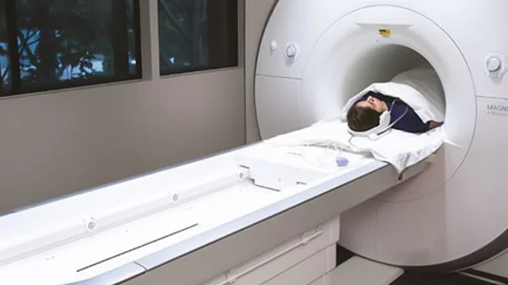
Cardiac MRI a powerful tool for predicting sudden cardiac death, arrhythmia after CIED implantation
The presence of myocardial fibrosis (MF) on cardiac MRI (CMR) can help anticipate adverse arrhythmic events, according to new findings published in the Journal of the American College of Cardiology.[1] This may offer a way to improve patient selection for cardiac implantable electronic devices (CIEDs).
The study’s authors noted that CIEDs are an unquestionably effective tool for preventing sudden cardiac death (SCD) and other significant events among certain high-risk patients. However, “only a minority” of patients ever directly benefit from receiving a CIED — and they put patients at a “lifelong risk” of serious complications.
The presence of MF, the authors wrote, has been linked to an increased risk of a ventricular arrhythmia. Could this finding help clinicians more accurately predict which patients may need a CIED — and which may not?
The CMR-CIED study included data from 700 patients who underwent CIED implantation from June 2002 to June 2018 at a single facility in the United Kingdom. The mean patient age was 68 years old, and CMR results from before the device was implanted was available for all patients. The median follow-up period was nearly 7 years.
Overall, SCD was seen 3.85% of patients. The study’s secondary endpoint — a composite of SCD, resuscitated cardiac arrest, appropriate ICD therapy and sustained ventricular tachycardia (VT) or ventricular fibrillation — was seen in 17.3% of patients.
“An SCD was defined as a natural, unexpected death caused by cardiac causes, heralded by an abrupt loss of consciousness within one hour of the onset of acute symptoms, in the absence of symptoms to suggest an acute coronary event,” wrote first author Francisco Leyva, MD, a cardiology professor and cardiac device therapy specialist out of Aston University in the United Kingdom. “Sustained VT was defined as a ventricular rhythm faster than 100 beats/min lasting at least 30 seconds or requiring termination caused by hemodynamic instability or by antitachycardia pacing or shocks.”
Visually apparent MF was associated with an elevated risk of SCD. All patients who experienced SCD showed apparent MF in their CMR results from prior to implantation, meaning the negative predictive value was 100%. Also, the absence of apparent MF “virtually excluded” the study’s secondary endpoint, resulting in a negative predictive value of 99%.
Left ventricular ejection fraction, the team added, was unable to accurately anticipate either SCD or the secondary endpoint.
“These findings suggest that, in patients who meet current guideline criteria for CIEDs, a defibrillator capability may only be required in patients with MF on visual assessment at a preimplantation CMR,” the authors wrote.
Related Cardiac Imaging Content:
Cardiac MRI sheds new light on vaccine-related myocarditis
TEE improves 30-day outcomes for patients undergoing cardiac valve or proximal aortic surgery
FDA clears AI-powered cardiac platform for pediatric patients
Reference:

