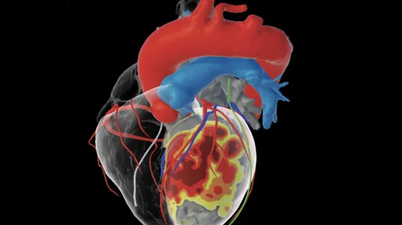
3D cardiac modeling solution for VT ablation receives FDA clearance
inHEART Medical, an international healthcare company with offices in France and the United States, has received clearance from the U.S. Food and Drug Administration (FDA) for its new 3D visualization software designed to improve the management of ventricular tachycardia (VT) ablation and other cardiac ablation procedures in the electrophysiology (EP) lab.
The software solution uses a proprietary algorithm to evaluate CT and MRI results and then build an interactive 3D model of the patient’s heart. Physicians can then reference those models before and during cardiac procedures.
According to data presented at European Heart Rhythm Association (EHRA) 2021, using this new software can lead to significant reductions in VT procedure times and higher success rates.
“We have been using the inHEART solution in clinical research for several years. Now with its commercial availability, inHEART will become part of our standard of care for planning and guiding therapeutic interventions,” Jeffrey Winterfield, MD, arrhythmia service chief and cardiac electrophysiology chair at MUSC Health, said in a prepared statement from inHEART. “The detailed substrate information in the 3D models allows us to pinpoint with accuracy and precision the arrhythmogenic areas in the scar tissue and target our ablation strategy accordingly. This information is invaluable for simplifying and enhancing our approach to VT ablation therapy.”
“With this clearance, we will grow inHEART's presence in the U.S. as we work to improve patient care for long, complex cardiac arrhythmias,” added Todor Jeliaskov, president and CEO of inHEART. “Our goal is to make a VT ablation as straightforward as an atrial fibrillation ablation so that VT ablation is no longer limited to academic VT centers.”
Related Cardiac Modeling Content
VIDEO: HeartFlow FFR-CT sees increased interest after inclusion in the 2021 Chest Pain Guidelines
Harvard researchers develop 3D model of human ventricle
Worth the wait: After years of trying, researchers 3D print a working heart pump with human cells

