MRI in patients with abandoned pacemaker and ICD leads
Magnetic resonance imaging (MRI) can heat or cause some types of metal to migrate inside the body, including metal-based tattoo inks and medical implants.
MRI safety protocols call for a list of these possible safety threats to be made prior to a patient being scanned. Some patients may have abandoned leads, or fragments of leads, from pacemakers or implantable cardioverter defibrillators (ICD), potentially putting them at an increased risk of adverse events.
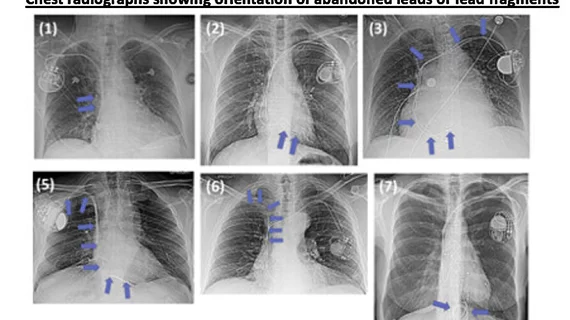
A recent study by electrophysiologists and radiologists at Banner University Medical Center and University of Arizona looked at the safety of these leads in real-world patients.[1]
“Patients with abandoned leads or lead fragments are considered at risk because of the unpredictable effects of lead tip heating during MRI and, as a secondary consideration, the risk associated with the CIED,” corresponding author Michael Morris, MD, with the department of radiology and division of cardiology at Banner University Medical Center Phoenix, and colleagues explain. “Although in vitro studies suggest that the length and orientation of fractured or abandoned leads contribute to heating during MRI, the effect in a clinical setting has not been reported. Additionally, it is unknown whether the length and orientation of abandoned leads or lead fragments affect CIED function in patients undergoing MRI.”
For the study, researchers identified 80 patients with 126 abandoned CIED leads or lead fragments who underwent 107 1.5T MRI examinations between March 2014 and July 2020.
CIED settings before and after MRI were reviewed, with clinically significant variations defined as a composite of the change in capture threshold of at least 50%, in sensing of at least 40%, or in lead impedance of at least 30% after MRI interrogation. Adverse clinical events were assessed at MRI and up to 30 days after. Univariable and multivariable analysis was performed.
The researchers said there were no reported deaths, clinically significant arrhythmias, or adverse clinical events within 30 days of MRI.
However, three patients with abandoned leads had a significant change in the composite of capture threshold, sensing or lead impedance. In a multivariable generalized estimating equation analysis, lead orientation, lead length, MRI type, and MRI duration were not associated with a significant change in the composite outcome. This included several patients that had lead wires in various shapes and positions that researchers watched closely to see if shape or position caused any adverse effects.
The group concluded MRI in the presence of abandoned CIED leads or lead fragments of varying lengths and orientations was not associated with adverse clinical events.
The leads that connect the heart to CIEDs wear out over time or are replaced with new wires when there is a device change procedure when battery levels run low, or if a device malfunctions or becomes infected. The standard of care up until recent years was to abandon the lead because of safety concerns related to removing the leads that become encased in scar tissue inside the veins where they are implanted. Many centers now remove these leads to prevent complications such as venous occlusion, or to ensure there is room for new leads, especially in younger patients who may undergo numerous can replacements over several decades.

