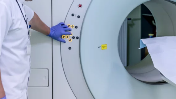
Study suggests MRI should be standard practice for NSTEMI diagnosis, treatment
Bypassing standard angiography and skipping straight to an MRI might help physicians more easily identify and treat patients who have suffered non-ST segment elevation myocardial infarction (NSTEMI), a team at Weill Cornell Medicine and NewYork-Presbyterian have found.
John F. Heitner, MD, an associate professor of clinical medicine at Weill, and colleagues said in Circulation this month that it can be difficult for physicians to pinpoint the artery that caused a patient’s heart attack, and their go-to for locating it tends to be coronary angiography. With angiography, an interventional cardiologist takes X-rays of a patient’s vessels during a cardiac catheterization, and MRI is only considered if angiography results aren’t clear to the cardiologist.
“We sought to learn whether an MRI performed first could improve clinicians’ ability to identify the affected artery,” Heitner said in a release. “Many patients with NSTEMI have diseases in several blood vessels or have no significant blockages at all that make it difficult to identify the correct artery that caused the heart attack. We suspected the MRI’s ability to directly visualize the heart muscle that was affected could improve accuracy, and our research suggests that to be the case.”
Heitner et al. recruited 114 patients for their study, all of whom had presented to one of three centers with their first heart attack. Participants underwent MRI followed by angiography, and images from both procedures were later reviewed independently and blindly to identify narrow or clogged arteries. The interventional cardiologist treating the patients was blinded to the MRI results.
The team found the artery in question wasn’t identifiable by angiography alone in 37% of cases. Of those cases, MRI was able to identify the correct artery 60% of the time and resulted in a new non-coronary artery disease (CAD) diagnosis 19% of the time. Even in patients whose angiography results were conclusive, MRI went on to identify a second affected artery in 14% of patients and a non-CAD diagnosis in 13%.
Overall, MRI led to a new diagnosis involving a damaged artery in 30% of cases and a diagnosis unrelated to the artery in 15% of cases.
“This study suggests that using an MRI should be the standard practice in NSTEMI patient diagnosis and care,” Heitner said. “We want to make sure that we are treating the artery that caused the heart attack, as this will likely lead to better long-term outcomes for the patient.”

