
New imaging technique captures human heart with 'unprecedented detail'
![Researchers with University College London and the European Synchrotron Radiation Facility (ESRF) have used a new X-ray technique, hierarchical phase-contrast tomography (HiP-CT), to capture images of the human heart in unprecedented detail. The group shared its images, as well as a full analysis, in Radiology.[1]](/sites/default/files/styles/top_stories/public/2024-07/screenshot_2024-07-17_at_3.40.36_pm.png.webp?itok=HKdrGjeC)
Researchers have used a new X-ray technique, hierarchical phase-contrast tomography (HiP-CT), to capture images of the human heart unlike any that have ever been seen before. The group shared the images, as well as a full analysis, in Radiology.[1]
“The atlas that we’ve created in this study is like having Google Earth for the human heart,” senior author Peter Lee, DPhil, a professor of materials science with the department of mechanical engineering at University College London, said in a statement. “It allows us to view the whole organ at global scale, then zoom in to street level to look at cardiovascular features in unprecedented detail.”
Lee et al. created two images of the human heart for the study, one from a 63-year-old donor with no known cardiovascular disease and another from an 87-year-old donor with a history of ischemic heart disease, hypertension and atrial fibrillation. The hearts had to be donated to gain this level of detail, researchers noted—the radiation required would be too high for a living person.
“One of the major advantages of this technique is that it achieves a full 3D view of the organ that’s around 25 times better than a clinical CT scanner,” Lee added. “In addition, it can zoom in to cellular level in selected areas, which is 250 times better, to achieve the same detail as we would through a microscope, but without cutting the sample. Being able to image whole organs like this reveals details and connections that were previously unknown.”
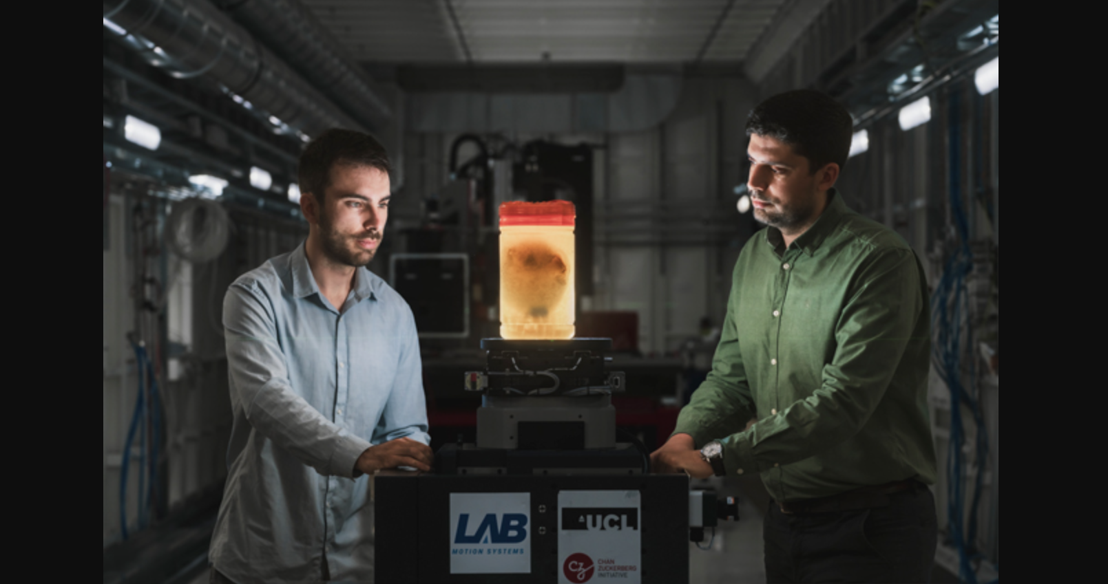
Imaging organs at the ESRF, the European Synchrotron, at BM18 beamline. Using an advanced synchrotron technique called HiP-CT, scientists have imaged, for the first time, two whole human hearts at 20-micron resolution, zooming in to 2-micron details in 3D. Image/caption courtesy of ESRF/Stef Cande.
Human hearts imaged at European Synchrotron Radiation Facility, 'world's brightest synchrotron light source'
The hearts were imaged using the BM18 beamline at the European Synchrotron Radiation Facility (ESRF) in France, known as the “world’s brightest synchrotron light source.” Multiple ESRF specialists served as co-authors on the Radiology study.
“BM18 is currently the only place in the world where complete human organs can be imaged with such a high level of contrast, and we are still quite far from the limits of the beamline capabilities,” co-author Paul Tafforeau, ESRF scientist, said in a separate statement. “The main limiting factor is the processing of the very large data produced by HiP-CT.”
What's next for University College London, European Synchrotron Radiation Facility?
The groups involved in producing these images hope their work can help cardiologists learn more about the different ways cardiovascular disease and other cardiac condition impact the human heart.
“We believe that our findings will help researchers understand the onset of cardiac rhythm abnormalities and also the efficacy of ablation strategies to cure them,” co-author Andrew C. Cook, a heart anatomist with University College London, explained. “For example, we now have a way to determine differences in the thickness of tissue and fat layers located between the outer surface of the heart and the protective sac surrounding the heart, which could be relevant when treating arrhythmia.”
The team also noted that many more hearts still need to be imaged using this advanced technology—these initial images represent just the beginning.
Click here to read the full analysis in Radiology.

Michael has more than 19 years of experience as a professional writer and editor. He has written at length about cardiology, radiology, artificial intelligence and other key healthcare topics.