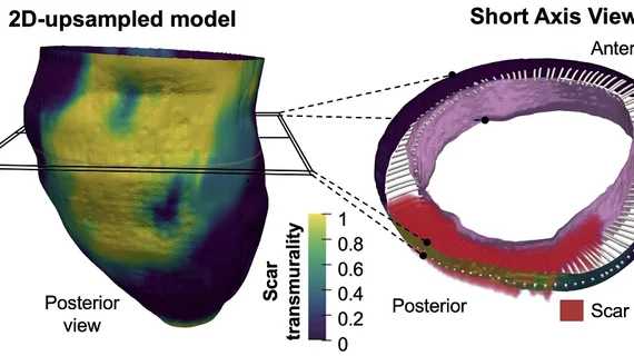
New imaging technique maps scar tissue after a heart attack
Researchers have developed a new technique for identifying the 3D features of scar tissue formed after a myocardial infarction, sharing their findings in Scientific Reports.
The method, which involves “3D transmural mapping of scar tissue in the infarcted muscle,” was designed so that is fully compatible with standard cardiac magnetic resonance (CMR) sequences. In addition, it only requires conventional 2D delayed gadolinium-enhanced CMR; no 3D CMR studies are required.
“This approach can thus significantly shorten the time needed for image acquisition, easing access to in-demand nuclear magnetic resonance scanners,” senior author David Filgueiras-Rama, a researcher with the Spanish National Center for Cardiovascular Research in Madrid, Spain, said in a statement.
The team noted that their new technique helped them pinpoint multiple key findings. For example, low scar transmurality on CMR is associated the frequency of ventricular tachycardias. Also, patients with low scar transmurality are more likely to have ventricular tachycardia recurrences later on during follow-up examinations.
“This novel methodology may provide an efficient approach in clinical practice after manual or automatic segmentation of myocardial borders in a small number of conventional 2D slices and automatic scar detection,” Filgueiras-Rama et al. wrote.
Read the full Scientific Reports analysis here.

