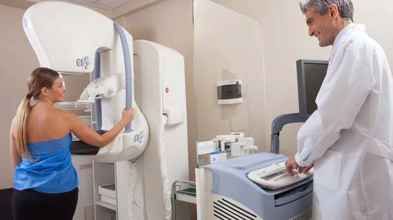
What routine mammograms can tell us about a woman’s CVD risk
Signs of breast arterial calcification (BAC) on a patient’s routine mammogram is associated with a heightened risk of cardiovascular disease (CVD), according to new research published in Circulation: Cardiovascular Imaging.[1]
“Among asymptomatic women, the first manifestation of underlying coronary heart disease is often unexpected acute myocardial infarction or sudden death,” wrote lead author Carlos Iribarren, MD, MPH, PhD, a specialist with the Kaiser Permanente Northern California Division of Research, and colleagues. “Furthermore, although women tend to have lower burden of obstructive coronary artery disease on angiography, they tend to have worse prognosis after myocardial infarction compared with men. Thus, a further investigation of sex-specific CVD risk markers is imperative.”
Iribarren and colleagues examined data from more than 5,000 women who participated in the MINERVA study, a massive collection of screening mammograms from a diverse group of postmenopausal women. All participants were between the ages of 60 and 79 years old at the time of their mammogram and had no prior history of CVD or breast cancer. They all received care at one of nine facilities in the same U.S. health system from October 2012 to February 2015.
Participants were followed for approximately 6.5 years after the mammogram.
Overall, signs of BAC were seen in the screening mammogram results of 26.5% of patients. Patients with BAC tended to be older and were more likely to be white or Hispanic. They were also more likely to have pre-diabetes, diabetes or hypertension.
Women with BAC were also 51% more likely to develop heart disease or stroke and 23% more likely to develop any type of CVD.
“Currently, it is not the standard of care for BAC visible on mammograms to be reported,” Iribarren said in a statement. “Some radiologists do include this information on their mammography reports, but it’s not required. We hope that our study will encourage an update of the guidelines for reporting breast arterial calcification from routine mammograms. Our study has moved the needle toward recommending routine assessment and reporting of breast arterial calcification in postmenopausal women.”
Taking BAC findings into consideration when developing risk calculators related to CVD, Iribarren added, could “help improve cardiovascular risk reduction strategies.”
Sadiya S. Khan, MD, and Natalie A. Cameron, MD, two specialists with the Northwestern University Feinberg School of Medicine in Chicago, wrote a separate editorial focused on this analysis for Circulation: Cardiovascular Imaging.[2]
“Iribarren et al.’s study is an important addition to a large body of literature examining the promise of imaging markers for early detection of subclinical CVD to guide prevention efforts,” the two co-authors wrote.
In a prepared statement, Khan added that BAC “may suggest poor heart health,” but it isn’t the only marker clinicians should consider.
“It is really important to note that the absence of BAC did not translate into low risk and should not be falsely reassuring when no breast arterial calcification is present,” she said. “Optimal risk factor control is equally important for all women with and without breast arterial calcification.”
Related Cardiac Imaging Content:
AI-powered ECG analysis could boost care for patients with hypertrophic cardiomyopathy
AFib, AI and heart-healthy diets: European Society of Cardiology previews EHRA 2022
CT a low-risk imaging alternative to invasive coronary angiography for suspected CAD
Cardiac MRI a powerful tool for predicting sudden cardiac death, arrhythmia after CIED implantation
References:

