World’s smallest endoscope could make a big difference in fight against heart disease
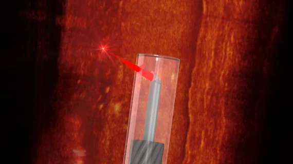
Researchers have developed what they believe is the world’s smallest imaging device, a microscopic endoscope capable of scanning the inside of blood vessels.
Sharing its findings in Light: Science & Applications, the team noted that this new device can “allow safe access to delicate but difficult-to-reach organs, enable a high-resolution cross-sectional imaging capability, and potentially lead to enhanced patient safety and improved health outcomes.”
The device, less than 0.5 millimeters across, was crafted using 3D printing technology. The researchers see significant potential in its ability to help treat patients struggling with heart disease.
“A major factor in heart disease is the plaques, made up of fats, cholesterol and other substances that build up in the vessel walls,” lead author Dr. Jiawen Li, faculty of health and medical sciences at the University of Adelaide in Australia, said in a statement. “Preclinical and clinical diagnostics increasingly rely on visualizing the structure of the blood vessels to better understand the disease. Miniaturized endoscopes, which act like tiny cameras, allow doctors to see how these plaques form and explore new ways to treat them.”
Simon Thiele, MSc, a co-author of the study from the University of Stuttgart, said modern technology made the team’s work possible.
“Until now, we couldn’t make high quality endoscopes this small,” he said. “Using 3D micro-printing, we are able to print complicated lenses that are too small to see with the naked eye.”
The full Light: Science & Applications study is available here.

