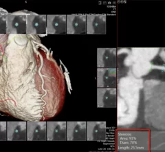
Cardiac Imaging

While cardiac ultrasound is the widely used imaging modality for heart assessments, computed tomography (CT), magnetic resonance imaging (MRI) and nuclear imaging are also used and are often complimentary, each offering specific details about the heart other modalities cannot. For this reason the clinical question being asked often determines the imaging test that will be used.

Patients and technologists benefit from reduced dose of UltraSPECT image reconstruction software

UltraSPECT, a leading provider of nuclear medicine (NM) image reconstruction technology that reduces radiopharmaceutical dose and acquisition time, announces today the installation of its proprietary Wide Beam Reconstruction (WBR™) software at nearly ten healthcare facilities.

MR helps predict cerebrovascular disease

Phase wave velocity (PWV) from the aortic arch obtained with phase-contrast MR imaging may prove a useful tool for determining the risk of cerebrovascular disease. In a study published in the June issue of Radiology, PWV was an independent predictor of subsequent white matter hyperintensity volume. 

Do men's and women's hearts burn fuel differently?

Researchers at the University of Illinois at Chicago College of Medicine will study gender differences in how the heart uses and stores fat -- its main energy source -- and how changes in fat metabolism play a role in heart disease, under a new $2 million, 4-year grant from the National Heart, Lung and Blood Institute.

App outperforms email for sending ECG images

An experimental cellphone app that transferred echocardiogram (ECG) images from emergency personnel to hospitals proved to be faster and more reliable than emailing images, according to an oral abstract presented May 17 at the American Heart Association’s Quality of Care and Outcomes Research scientific sessions in Baltimore.

Society of Cardiovascular Computed Tomography Announces Finalists for the 7th Annual Toshiba Young Investigator Award

The Society of Cardiovascular Computed Tomography (SCCT) announces the finalists for the 7th Annual Toshiba Young Investigator Award (YIA).  Sponsored by an educational grant from Toshiba America Medical Systems, Inc., the YIA program supports the professional and clinical development of top radiology residents and cardiology fellows within five years of completing a training program.   

One-stop CT, MR imaging: Future in diabetic care?

Quantifying body fat and other clinical factors in obese patients with diabetes using CT or magnetic resonance (MR) may help physicians detect and manage comorbidities such as cardiovascular disease, according to a review published in the May issue of Diabetes Technology & Therapeutics. But is that enough to justify “one-stop shop” imaging?

Beaumont cardiologist Kavitha Chinnaiyan, M.D., receives excellence in research award

Kavitha Chinnaiyan, M.D., director of Advanced Cardiac Imaging Education at Beaumont Hospital, Royal Oak, has received the Frank J. McDevitt, D.O., Award for Excellence in Health Policy Research for Physicians.

The Heart Rhythm Society announces Dr. Hugh Calkins as the organization’s 35th president

Hugh Calkins, MD, FHRS, CCDS, was named the president of the Heart Rhythm Society (HRS) during the 34th Annual Scientific Sessions, held in Denver last week.  Announced during the Presidents’ Reception on Friday, May 10th, Dr. Calkins now serves as the 35th president of HRS, making Anne M. Gillis, MD, FHRS, the immediate past president. Dr. Calkins is currently a Nicholas J. Fortuin Professor of Cardiology and Professor of Medicine at the Johns Hopkins University School of Medicine in Baltimore, Maryland.  

Around the web

Several key trends were evident at the Radiological Society of North America 2024 meeting, including new CT and MR technology and evolving adoption of artificial intelligence.

Ron Blankstein, MD, professor of radiology, Harvard Medical School, explains the use of artificial intelligence to detect heart disease in non-cardiac CT exams.