Cardiac Imaging

While cardiac ultrasound is the widely used imaging modality for heart assessments, computed tomography (CT), magnetic resonance imaging (MRI) and nuclear imaging are also used and are often complimentary, each offering specific details about the heart other modalities cannot. For this reason the clinical question being asked often determines the imaging test that will be used.

St. Jude launches 3D nav system for vascular, cardiac anatomy

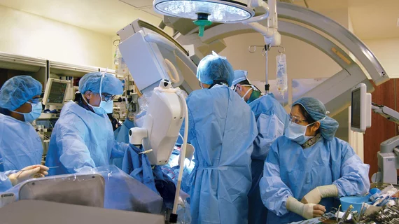
St. Jude Medical has launched its MediGuide Technology, a 3D navigation system intended for the evaluation of vascular and cardiac anatomy on a recorded fluoroscopic image instead of live fluoroscopy (a series of x-ray images).

Obese patients should have MPI using conventional SPECT camera

Patients with a body mass index of 40 kg/m2 or greater should be scheduled for myocardial perfusion imagingon a conventional SPECT camera, as it is difficult to obtain diagnostic image quality on a cadmium-zinc-telluride camera, according to a study in the September issue of the Journal of Nuclear Medicine.

CTA in reoperative CABG is fiscally justified

The use of CT angiography (CTA) to help guide preventive surgical strategies for patients undergoing redo CABG appears to be justified, based on recently published results.

Carotid Conundrum: Role of Ultrasound Screening

Despite recommendations to the contrary, screening for carotid artery stenosis remains prevalent. Check out the pros and cons.

Overnight subspecialty coverage pushes CT use higher in community EDs

Overnight imaging subspecialty coverageas opposed to a more traditional model of on-call preliminary interpretations by a residentresulted in an increase in CT utilization in community emergency departments (EDs), according to a study published in the September issue of the American Journal of Roentgenology.

ESC: Ultra-fast CT scanner put to test for coronary blockages

A 320-detector CT scanner can accurately identify which people with chest pain need, or dont need, an invasive procedure such as cardiac angioplasty or bypass surgery to restore blood flow to the heart, according to the results of the CORE 320 study presented Aug. 28 at the European Society of Cardiology Congress in Munich.

ESC: DeFACTO misses mark but shows promise for diagnosing CAD

DeFACTO, a study designed to assess the performance of a noninvasive method for gauging the physiologic significance of coronary artery disease (CAD), failed to meet its prespecified primary endpoint, researchers reported Aug. 26 at the European Society of Cardiology Congress in Munich. But that may be the result of the studys high standards, according to the author of an editorial that accompanied the studys simultaneous publication online in the Journal of the American Medical Association, who agreed with the researchers that the approach holds promise.

CAC imaging bests other markers for intermediate-risk patients

Coronary artery calcium (CAC) provided superior discrimination and risk reclassification compared with other risk markersankle-brachial index, high-sensitivity C-reactive protein and family historyall of which are independent predictors of incident coronary heart disease/cardiovascular disease in intermediate-risk individuals, according to a study in the Aug. 22/29 issue of the Archives of Internal Medicine.

Around the web

Several key trends were evident at the Radiological Society of North America 2024 meeting, including new CT and MR technology and evolving adoption of artificial intelligence.

Ron Blankstein, MD, professor of radiology, Harvard Medical School, explains the use of artificial intelligence to detect heart disease in non-cardiac CT exams.