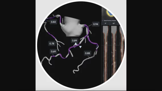
Cardiac Imaging

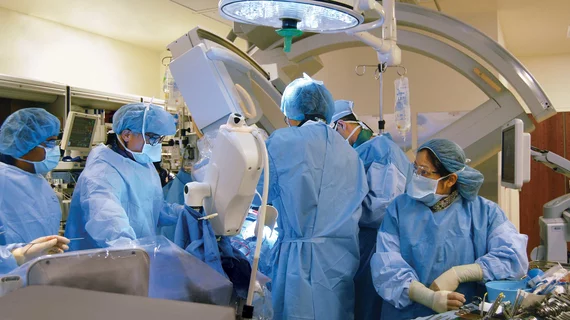
While cardiac ultrasound is the widely used imaging modality for heart assessments, computed tomography (CT), magnetic resonance imaging (MRI) and nuclear imaging are also used and are often complimentary, each offering specific details about the heart other modalities cannot. For this reason the clinical question being asked often determines the imaging test that will be used.

The lost art of listeningHas bedside auscultation given way to imaging?

Police officers carry a badge. Carpenters have a tool belt. Doctors have stethoscopes. Children putting together Halloween costumes intuitively understand that certain objects symbolize certain professions. But in the case of the stethoscope, is actual use declining?

Siemens expands Acuson line of ultrasound systems

Siemens Healthcare has released the Acuson S1000 ultrasound system.

ASNC Releases Dose-Reduction Guidelines for Nuclear Cardiology

Technological improvements in image acquisition and software processing in nuclear cardiology should allow physicians to shave patient imaging times dramatically or cut radiation doses fourfold, according to a new preferred practice statement from the American Society of Nuclear Cardiology (ASNC).

Feature: PCI, medical therapy for stable angina face off

While it is widely known from literature that PCI is beneficial in reducing mortality and MI in patients with acute coronary syndrome, the role of PCI in treating stable coronary artery disease (CAD) remains controversial. A new meta-analysis has found that PCI for stable CAD patients had no benefit compared with initial medical therapy. In interviews, three researchers discuss the implications of that and other analyses.

Study: ED CT protocol linked with excess rad exposure

The introduction of a whole-body panscan CT protocol for blunt trauma in the emergency department (ED) raised the proportion of patients exposed to more than 20 mSv of radiation by 8 percent, according to a study published in the February issue of Emergency Medicine Australasia.

JACC: CT can help prevent undersizing of aortic valves

Multidetector CT (MDCT) 3D aortic annular dimensions are predictive of valve leakage following transcatheter aortic valve replacement (TAVR), according to a study published online Feb. 22 in the Journal of the American College of Cardiology.

JACC: CT trumps 2D echo for valve replacement

There could be a new gold standard for aortic annular evaluation before transcatheter aortic valve replacement (TAVR) as 3D CT imaging has been shown to be superior to 2D transesophageal echocardiography (TEE), according to a study published Feb. 22 online in the Journal of the American College of Cardiology.

Mixed bag for CT angiography

Findings on CT angiography (CTA) dominated two research journals in recent weeks, or so it would seem. Both Radiology and the Journal of the American College of Cardiology published several studies in the past month that scrutinized the use of CTA in the clinical setting.

Around the web

Several key trends were evident at the Radiological Society of North America 2024 meeting, including new CT and MR technology and evolving adoption of artificial intelligence.

Ron Blankstein, MD, professor of radiology, Harvard Medical School, explains the use of artificial intelligence to detect heart disease in non-cardiac CT exams.