Spectacular science: 3D model offers breathtaking look at what happens during a heart attack
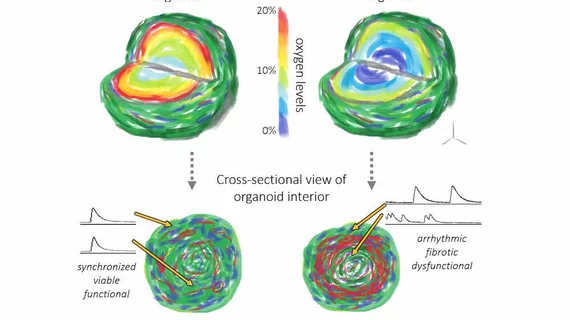
So what, exactly, occurs inside the human heart during a myocardial infarction? Researchers have developed 3D multicellular tissue samples that mimic what happens in eye-opening detail, publishing their research in Nature Biomedical Engineering.
These cardiac organoids are incredibly small—less than 1 millimeter in diameter, to be precise—but they could teach scientists a great deal about the cardiovascular health of the human species. Lead author Dylan Richards, PhD, of the bioengineering department at Clemson University, noted in a news release from the Medical University of South Carolina that cell samples or animal models are typically used for this type of research—but these new organoids paint a much more accurate picture.
“The hearts of rats and mice beat five to 10 times faster than those of humans,” Richards said. “How those mechanisms work physically—the electrophysiology and the pumping action—is just different because of the scale.”
Richards added that these organoids can improve what we know about cells respond in the immediate aftermath of a MI. Scientists typically view the human heart long after the damage is done. The breakthrough may also improve what we know about the impact of different medications.
“It could help us determine whether a drug is effective at preventing some of this damage or preventing a detrimental response to an oxygen shortage,” Richards said.
The full study from Nature Biomedical Engineering can be found here.

