Magnetocardiography IDs chest pain in 90 seconds
A 90-second chest scan could be a game-changer for triaging patients who present to the emergency department with undiagnosed chest pain, according to research out of Detroit.
Writing in the International Journal of Cardiology: Heart and Vasculature, lead author Margarita Pena, MD, and colleagues at Ascension St. John Hospital at Wayne State University School of Medicine said chest pain is one of the most common complaints in the ED—so common, in fact, that it’s the reason some 8 to 10 million Americans make a trip to the emergency room each year. And in such a high-stakes environment, it can be difficult to discern which cases are cardiac-related and need immediate attention.
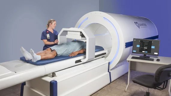
On Jan. 8, Ohio-based imaging tech company Genetesis, Inc., published a pilot study outlining the efficacy of its CardioFlux FAC Magnetocardiography (MCG), a noninvasive imaging device designed to measure and visualize the electromagnetic function of the heart. According to Pena et al.’s research, a 90-second MCG scan with the CardioFlux helped physicians determine a patient’s odds of experiencing an acute coronary syndrome.
Although only a small percentage of ED patients will be diagnosed with an ACS on any given day, Pena and colleagues said most people who present to the hospital with undiagnosed chest pain will be placed in an observation unit for extra tests and monitoring. The idea is that it’s better to be safe than sorry, but additional stays can rack up patients’ hospital bills and lead to pricey, unnecessary downstream tests that expose them to radiation and risky pharmaceuticals.
CardioFlux scans are performed at rest and don’t use any radiation or pharmaceuticals. The scanner studies the electrical activity in a patient’s myocardium and records a corresponding magnetic field map. Changes in that map are known to correspond with cardiac ischemia.
“Having a noninvasive diagnostic test that can be performed rapidly with almost no patient preparation in the emergency department observation unit or possibly incorporated into the ER workflow would be a game-changer in the evaluation of ED patients presenting with chest pain,” Pena said in a release. “This has a lot of potential to screen patients [for] coronary artery disease, not to mention the benefits of avoiding risks to the patient associated with radiation, adverse reactions to pharmacologic and contrast agents, as well as risks associated with hospitalization.”
For their study, Pena et al. administered a 90-second CardioFlux MCG to 101 low- to intermediate-risk chest pain patients. The CardioFlux performed similarly to stress testing and coronary angiography with a demonstrated specificity of 78.3% and a negative predictive value of 92.3%.
“Now they’re looking at the high-risk population of patients that get [catheterized] so that we can see how this compares to catheterization,” Pena said.

