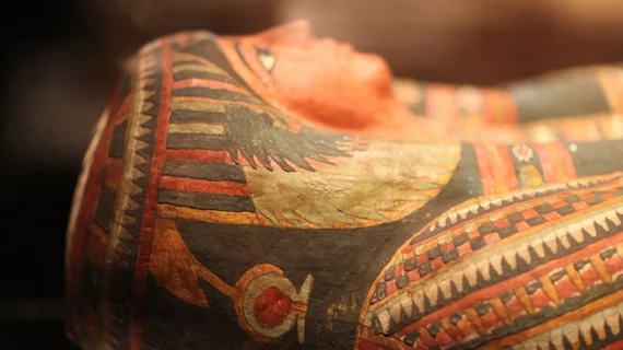
Cardiologists ID signs of widespread heart disease in ancient mummies
A team of cardiologists has used cardiac imaging technology to confirm that cardiovascular disease was a significant issue thousands of years ago, presenting its findings in European Heart Journal.[1]
The group used computed tomography (CT) to evaluate a total of 237 adult mummies from all over the world, including Egypt and ancient Peru. Signs of definite or probably atherosclerosis were identified in 37.6% of the samples.
“We found atherosclerosis in all time periods—dating before 2,500 BCE—in both men and women, in all seven cultures that were studied, and in both elites and non-elites,” lead author Randall Thompson, MD, a cardiologist at Saint Luke’s Mid America Heart Institute, said in a statement. “This further supports our previous observation that it is not just a modern condition caused by our modern lifestyles.”
The most involved vascular bed when it came to definite or probable atherosclerosis was the aorta (21.5%), following by the ilio-femoral (20.7%), popliteal-tibial (16%), carotid (14%) and coronary (0.4%) arteries.
A: Volume-rendered computed tomography image demonstrating extensive atherosclerosis (arrows) in the aorta of a female mummy from ancient Peru (Rosita). B: Multiplanar reconstruction: sagittal view computed tomography scan image demonstrates heavy calcific disease in the left carotid bulb (arrow). C: Thick slab maximum intensity projection: modified coronal computed tomography scan image demonstrates heavy coronary artery calcium, both in a female Egyptian mummy from the late Middle Kingdom—Second Intermediate Period. D: Number of mummies with none, mild to moderate (one to two vascular bed involvement), and severe (three to five vascular bed involvement) atherosclerotic calcifications for each of 13 epochs. Images and caption courtesy of Thompson et al., European Heart Journal.
Mummies included in the study came from cultures spanning more than 4,000 years. The estimated mean age was 40 years old—young by modern standards, but much older for the time. The researchers highlighted what today’s patients should take from these ancient samples.
“This study indicates modern cardiovascular risk factors—such as smoking, sedentary lifestyle, and poor diet—on top of the underlying, inherent risk natural to the human aging process may increase the extent and impact of atherosclerosis,” Thompson said. “This is why it is all the more important to control the risk factors we can control.”
The group also noted that it was very conservative with its estimates due to the risk that findings would be impacted by distorted tissue samples. In addition, a majority of the mummies only had a limited number of vascular beds that were able to be included in the analysis.
Even with these limitations, however, the authors believe these findings show that atherosclerosis has been prevalent for much longer than many cardiologists may realize.
Thompson has been involved in several past CT studies of ancient mummies from around the word.
Click here to read the full study in European Heart Journal, a publication of the European Society of Cardiology.

![A team of cardiologists has used cardiac imaging technology to confirm that cardiovascular disease was a significant issue thousands of years ago, presenting its findings in European Heart Journal.[1]](/sites/default/files/styles/no_crop/public/2024-05/screen_shot_2024-05-29_at_3.50.15_pm.png.webp?itok=xcN9Y0tz)
