
Harvard researchers unlock the secret to building 3D-printed heart muscles that can beat
Researchers at Harvard University have found that a new hydrogel ink, infused with gelatin fibers, makes it possible to 3D print a functional heart ventricle that beats like a human heart. The group shared its findings in Nature Materials, noting that the same technique can be used to 3D print heart valves, dual-chambered hearts and more.[1]
“People have been trying to replicate organ structures and functions to test drug safety and efficacy as a way of predicting what might happen in the clinical setting,” first author Suji Choi, PhD, a research associate with the Harvard John A. Paulson School of Engineering and Applied Sciences (SEAS), said in a prepared statement.
Creating these complex structures with 3D printing alone has never been possible, Choi et al. explained—until now.
Behind the technology
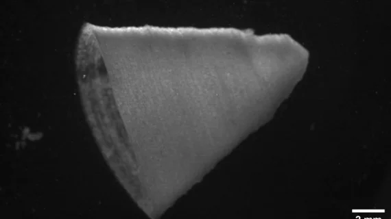
The fiber-infused gel (FIG) ink used to print these functional heart ventricles was created with a rotary jet spinning technique first developed in the lab of Harvard professor Kevin “Kit” Parker, PhD. Parker credits Luke MacQueen, PhD, a co-author of the Nature Materials study, with first proposing the idea of adding gelatin fibers to ink for this purpose.
“When Luke developed this concept, the vision was to broaden the range of spatial scales that could be printed with 3D printers by dropping the bottom out of the lower limits, taking it down to the nanometer scale,” Parker explained in the Harvard statement. “The advantage of producing the fibers with rotary jet spinning rather than electrospinning is that we can use proteins that would otherwise be degraded by the electrical fields in electrospinning.”
Choi used MacQueen’s concept to produce a sheet of material that was then broken into tiny fibers using sound waves. These fibers were then added to hydrogel ink, which was used to print the team’s functional heart ventricles.
“Compared to the real heart, our ventricle model is simplified and miniaturized,” Choi said, noting that her team is already working on future models that more closely resemble an actual human heart.
As the Harvard researchers push forward, they hope to learn more about the potential of FIG ink—but they also know they can’t limit their efforts to just one process.
“The goal is not to be tool driven—we are tool agnostic in our search for a better way to build biology,” Parker said.
Read the full study here.

Michael has more than 18 years of experience as a professional writer and editor. He has written at length about cardiology, radiology, artificial intelligence and other key healthcare topics.