What CV research looks like in space
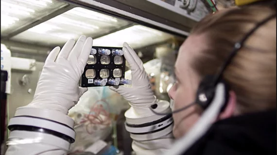
Researchers on the International Space Station are leveraging the microgravity conditions within the ISS U.S. National Laboratory to study heart cells in ways we cannot be conducted on the ground.
In an August 12 blog post, the ISS National Lab described the ongoing work of three scientists: Mary Kearns-Jonker, Arun Sharma and Chunhui Xu. The trio have all made or will make the journey to space, using ISS resources to study the effects of microgravity on cardiomyocytes.
Kearns-Jonker’s work centers around microgravity’s effects on human CV progenitor cells (CPCs)—immature heart cells that can be differentiated to represent several different types of cardiovascular cells—according to the post. The Loma Linda University researcher and her team found microgravity induces changes in CPCs that might be beneficial to ongoing research on Earth, which is focused on the potential use of CPCs in the development of regenerative therapies.
Sharma, a postdoctoral research fellow at Harvard, led the charge on a project that investigated how microgravity affected the function of cardiomyocytes derived from human induced pluripotent stem cells (iPSCs).
“We’re doing a variety of analyses to see how the heart cells beat differently or how their DNA changes due to microgravity,” he said in the blog post. “A big unanswered question is, what happens at the cellular level due to microgravity? And then, how can we use this new understanding to help cardiac patients here on Earth?”
Xu, of Emory University, and her team are gearing up for an upcoming investigation on the ISS that will analyze the ability of cardiomyocytes to differentiate from iPSCs and proliferate in microgravity. Xu et al.’s ground-based research found simulated microgravity increased the yield, purity and survival of such cardiomyocytes, and Xu said more realistic conditions are expected to enhance those effects.
“Large numbers of cardiomyocytes need to be generated in a more efficient way than currently possible,” she said. “When we combined simulated microgravity with 3D culturing techniques, we saw promising steps toward reaching that goal.”
Read more about the scientists’ work here.

