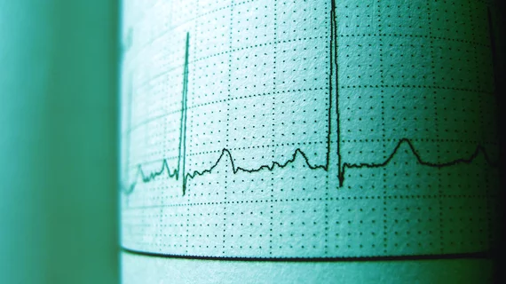
AI models capable of identifying RV, LV dysfunction in ECGs
Researchers have developed deep learning models capable of identifying right ventricular (RV) and left ventricular (LV) dysfunction from electrocardiogram (ECG) findings, sharing their findings in JACC: Cardiovascular Imaging.[1]
“The ECG is a cardinal investigation in the practice of cardiology,” wrote lead author Akhil Vaid, MD, a specialist with the Icahn School of Medicine at Mount Sinai in New York City, and colleagues. “It is ubiquitous, inexpensive, and is often the first investigation performed in emergency situations. However, it has an upper bound of usefulness secondary to its skill requirement and subjectivity. In addition, clinicians cannot identify subtle patterns that might indicate subclinical pathology, especially for conditions for which there are no interpretation guidelines.”
Vaid et al. developed their advanced artificial intelligence (AI) algorithms using data from a high-volume health system, focusing on more than 147,000 patients who received care from 2003 to 2020. All patients underwent echocardiography (echo) and an ECG. While more than 109,000 patients presented with a LV ejection fraction (LVEF) higher than 50%, more than 17,000 patients had an LVEF of 40% or less. The remaining patients — nearly 20,000 — fell in between those two measurements. The median age of each group was between 65 and 67 years old, and all groups were at least 50% male.
A machine-learning model was built that could classify a patient’s LVEF as ≤40%, >40% and ≤50% or >50%. The group also used natural language processing (NLP) to extract RV size and systolic function data from each patient’s imaging results and electronic health record (EHR) data. NLP was also used to identify signs of RV systolic dysfunction (RVSD) from a patient’s ECG.
The LVEF classification model had an area under the ROC curve (AUC) of 0.94 for identifying an LVEF of ≤40%, an AUC of 0.82 for identifying >40% and ≤50% and an AUC of 0.89 for identifying an LVEF of >50%. External validation resulted in AUCs of 0.94, 0.73 and 0.87, respectively.
In addition, the team’s AI models achieved an AUC of 0.84 when it came to predicting the study’s composite RV outcome. The AUC was the same for their internal testing and external validation.
Overall, the researchers wrote, their findings show that it is possible to develop, evaluate and externally validate deep learning models that can evaluate a patient’s left and right ventricles. Also, they added, they successfully created “an accurate NLP pipeline of outcomes from free-text echo reports”
“Our decision to not stratify RV disease according to severity was made to allow for early detection of disease,” the authors added. “Depending on clinical context, this may be adjusted to more severe disease. We posit performance in this context will also increase because there is a greater difference between normal and diseased cases.”
Related Heart Failure Content:
COVID-19 vaccines safe for patients with a history of heart damage
PCI boosts survival for ischemic HF patients with moderate-to-severe mitral regurgitation
Cancer, cancer-related death much more common among heart failure patients
Telehealth provided value for heart failure patients during COVID-19 pandemic
Reference:

