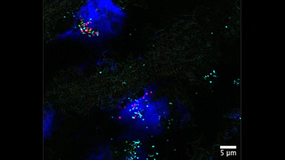
Up close and personal: Detailed cellular map of the heart offers ‘a goldmine of information’
A new cellular and molecular map of the human heart provides never-before-seen details that could make a big impact on patient care.
An international research team collaborated on the project, sharing its work in Nature. The group analyzed nearly 500,000 individual cells, completing what they are calling “the most extensive draft cell atlas to date of the human heart.”
The map is so detailed, in fact, that it can predict how various cardiac cells will communicate with one another at any given moment.
The ultimate goal is to use this map—part of the ongoing Human Cell Atlas initiative—to learn more about the heart and improve the treatment of a variety of cardiac ailments. For instance, the map’s detailed view of the heart’s blood vessels could help researchers learn new information about what they go through when a person has coronary heart disease.
“Millions of people are undergoing treatments for cardiovascular diseases,” co-senior author Christine E. Seidman, MD, a professor at Harvard Medical School and cardiovascular geneticist at Brigham and Women’s Hospital, said in a prepared statement. “Understanding the healthy heart will help us understand interactions between cell types and cell states that can allow lifelong function and how these differ in diseases. Ultimately, these fundamental insights may suggest specific targets that can lead to individualized therapies in the future, creating personalized medicines for heart disease and improving the effectiveness of treatments for each patient.”
“This project marks the beginning of new understandings into how the heart is built from single cells, many with different cell states,” added co-first author Daniel Reichart, MD, a research fellow in genetics at Harvard Medical School. “With knowledge of the regional differences throughout the heart, we can begin to consider the effects of age, exercise and disease and help push the field of cardiology toward the era of precision medicine.”
The team even examined one of the biggest topics facing cardiology today: COVID-19’s short-term, and long-term, impact on the heart.
“We mapped the cardiac cells that can be potentially infected by SARS-CoV-2 and found that specialized cells of the small blood vessels are also virus targets,” co-senior author Michela Noseda, a lecturer at Imperial College London, said in the same statement. “Our datasets are a goldmine of information to understand subtleties of heart disease.”
Click here to read more about this powerful map in Nature.

