A Step Closer to Precision Medicine? Gene Editing Process Leads to Personalized Advice for Heart Patient
University of Pennsylvania researchers have developed a stem cell–based test to determine whether genetic variants in heart muscle cells are benign or pathogenic. In other words, they’ve added some certainty to variants of uncertain significance (VUS)—the tricky alleles that can either contribute to the development of diseases or be completely harmless.
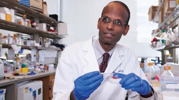
“Sometimes it’s worse to not have any idea whether it’s harmful than to know that it’s harmful and actually have some certainty to know that, ‘OK, I’m at risk and so there’s certain things I need to do,’” says Kiran Musunuru, MD, PhD, an associate professor of cardiovascular medicine and genetics at Penn.
Musunuru and colleagues believe they’ve made a breakthrough in taking the wealth of data generated from genome sequencing and turning it into actionable clinical information. The group reported the first use of a stem cell–based assay to inform the treatment of an actual patient in real time (Circulation 2018;138:2852-4).
Their platform allowed them to determine that a 65-year-old woman’s particular variant of TNNT2, a troponin-encoding protein that has been associated with cardiomyopathy, wasn’t pathogenic. The patient had been worried that her hypertrophic cardiomyopathy would be likely to appear in her two daughters and six grandchildren, but Musunuru and colleagues found her TNNT2 variant likely didn’t contribute to the disease. Based on this conclusion, they recommended her other family members not receive any extra genetic testing or screening for the time being, but continue to be monitored in the future for potential thickening of the heart wall.
“It’s a proof of concept, but the fact that we were able to apply it to an actual patient in the real world and actually have an appreciable effect on how we were managing her and the decision-making process about whether to test her children or not, that encouages us that this can actually be done with a lot more patients,” Musunuru told Cardiovascular Business.

Source: Musunuru Laboratory, University of Pennsylvania.
Ten-week process
The researchers modeled the patient’s heart cells by taking induced pluripotent stem cells, adding the TNNT2 genetic variant and then converting them in a petri dish into heart muscle cells.
They used isoproterenol—which is similar to adrenaline—to see if the heart cells would beat faster in the dish, as they would if a typical person were given a shot of adrenaline or a related substance. With harmful mutations of TNNT2, the cells no longer respond to isoproterenol, so a steady heart rate serves as an indicator that the allele contributes to the development of a cardiomyopathy.
In their patient’s case, the cardiomyocytes responded normally with faster beating, offering reassurance that the TNNT2 variant wasn’t contributing to her disease progression.
The whole process took 10 weeks— enough time for the team to give the patient a clear answer before her follow-up appointment. “It might seem like a long time, but if you think about all the things we do in modern medicine, it can take 10 weeks just to schedule a CT scan or an MRI after a visit with your physician,” Musunuru says. “We’re not talking years, we’re talking on the order of weeks, so there is that potential for the test to be done in enough time for it to have a meaningful impact. … It couldn’t be done in an emergency situation, obviously, but we’re talking about diseases that take a long time to develop.”
Musunuru says the cost of the process totaled “several thousand dollars,” which he points out is “not all that different” from diagnostic imaging procedures. As he and his colleagues continue to work with the platform, they hope it becomes more efficient and less costly.
Progressing toward precision medicine
The group also has used this technique to clarify a decision in patient care for a patient with a variant of LMNA, another gene that has been linked to cardiomyopathy. And they are using a similar pathway to test VUS in a gene related to familial hypercholesterolemia.
While the process has already proven capable of providing personalized information to patients and their families, Musunuru envisions an even more impactful application down the road. He says there are about 4,000 possible variants of TNNT2. He’d like to test them all over the next few years—“or however long it takes”—and develop a database on which ones are benign vs. pathogenic. Then, clinicians could immediately tell patients whether their variants are relevant or not, without taking the time or the expense to perform the testing process.
“I think that is the long-term promise of this,” Musunuru says. “It will take some years to do it, but when we do get to that point, we’ll be really doing what people talk about now as precision medicine. We’ll be able to highly personalize your genome sequence.”

