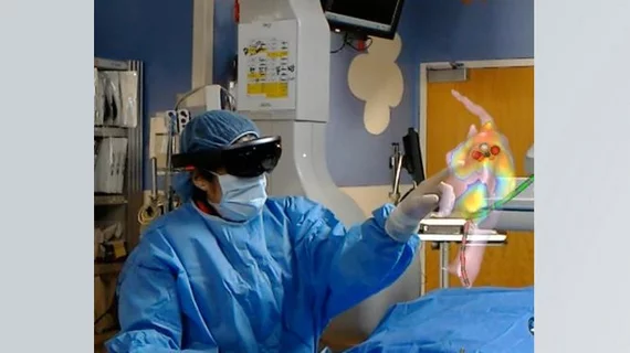
Meet ELVIS, the holographic display that improves cardiologist accuracy
ELVIS, it seems, has entered the operating room.
Using a new holographic display system in the operating room can improve physician accuracy during cardiac ablation procedures, according to new findings out of St. Louis.
Researchers combined custom software with Microsoft’s HoloLens technology to build the Enhanced Electrophysiology Visualization and Interaction System—or ELVIS for short. ELVIS produces 3D digital images of the inside of the patient’s heart, and they can be moved and manipulated by the cardiologist during the procedure.
The team tested the system on 16 pediatric patients at a single facility in St. Louis, publishing their research in both the Journal of the American College of Cardiology–Clinical Electrophysiology and IEEE Journal of Translational Engineering in Health and Medicine.
Physicians received a short training session before using the system and were tested on accuracy during a post-procedure waiting period. The system helped reduce the number of ablation lesions made outside of the target area from 34% all the way to down to 6%.
“We expect that this will improve patient outcomes and potentially reduce the need for repeat procedures,” co-author Jonathan R. Silva, department of biomedical engineering at Washington University in St. Louis, said in a statement.
“What ended up being equally important, if not more important, was that this was the springboard for everything that is to come, not only that we can visualize it better, but that we can control it,” added co-author Jennifer Silva, MD, an associate professor of pediatrics at the Washington University School of Medicine. “There are people working in this extended reality space who have come to conclusions that the control is the strongest value-add, particularly in medical applications.”
The researchers have licensed their technology to a vendor as development continues.
Click here to read the full study.

