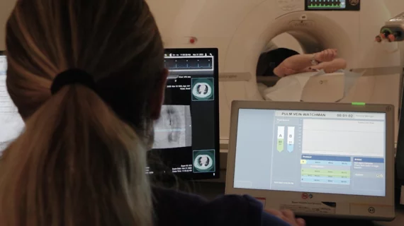
CCTA a helpful tool for interventional cardiologists planning coronary procedures
Coronary CT Angiography (CCTA) has evolved into a much more helpful resource than interventional cardiologists may have originally believed. In fact, the Society of Cardiovascular Computed Tomography (SCCT) emphasized in a new expert consensus statement that CCTA provides specialists with an effective tool in the preparation and optimization of coronary procedures.
The statement emphasizes that CCTA appears to be surpassing its place among interventional cardiologists as “a mere diagnostic tool.” Combining CCTA with fractional flow reserve (CT-FFR) or stress CT myocardial perfusion imaging (CT-MPI), the group wrote, can help clinicians assess a patient’s plaque burden, predict the success of revascularization, identify coronary lesions in need of additional treatment and make key decisions between percutaneous coronary intervention (PCI) and coronary artery bypass graft (CABG) surgery.
The statement’s authors also noted that CCTA is in many ways an upgrade compared to conventional invasive coronary angiography, providing multiple benefits that make it an especially promising heart teams for heart teams.
"It's quite analogous to using CT to plan a structural heart procedures, but still far too often our coronary interventional cardiology colleagues go into the cath lab without any information," Jonathon Leipsic, MD, FRCPC, MSCCT, chairman of the department of radiology at the University of British Columbia and a co-author of the statement, explained in a recent interview. "They know the patient has some symptoms and perhaps an equivocal stress test, but they don't know anything more than that, such as the extent of disease or what they are going to encounter. But given that the guidelines have shifted to a CT first strategy, more and more patients are going to have a CT and now it is about exploring how the CT can help plan coronary interventions to ensure more complete revascularization."
Leipsic said CT can bring not just anatomical information about the artery and the plaque, but also show vessel remodeling, offer information on plaque composition. It also offers functional information using virtual fractional flow reserve CT (FFR-CT), where FFR measurements can shown on a map off the entire coronary tree. He said this can help guide interventionists in planning without the need for using pressure wires in the cath lab. CT also can show the best viewpoints to see a specific coronary or lesion, which can help plan what angle the C-arm needs to be at to duplicate that view to save procedure time. CT also can enable stent sizing and give an idea of guide wires and other equipment needed prior to catheterizing the patient.
"If you go in to take out an appendix, you don't do an exploratory laparotomy, you do a CT," Leipsic explained. "You confirm if there is appendicitis and then come up with a plan. The same is true of tumor imaging, or abdominal aortic aneurisms. So now we have a rich anatomical dataset for the coronaries that gives you lesion length, stenosis morphology, and it gives us an understanding on the morphology of the plaque. And with FFR-CT, we now have physiology that can help you understand the depth of the abnormality, the pattern of the physiology."
The new expert consensus statement comes less than a year after cardiac CT was indicated as a front-line chest pain assessment tool in the 2021 chest pain guidelines published by members of the American Heart Association, American College of Cardiology, SCCT and several other industry organizations.[1]
The updated recommendation made a colossal impact on patient care, pushing interest in the modality throughout the United States to an all-time high.
The full SCCT statement was published in the Journal of Cardiovascular Computed Tomography, the official journal of the SCCT.[2] It was simultaneously published in EuroIntervention, the official journal of EuroPCR and the European Association of Percutaneous Cardiovascular Interventions.[3]

