Researchers receive $3.7M to attempt robotic heart surgery inside MRI scanner
Researchers from Case Western Reserve University have received a four-year, $3.7 million grant from the National Institutes of Health (NIH) to try and make a bit of history.
The group—which includes engineers, cardiologist, radiologists and other specialists—will attempt to perform a robotic-controlled left atrial appendage occlusion (LAAO) on a patient inside an MRI scanner; if successful, it would be the first procedure of its kind.
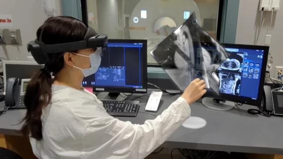
A clinician will perform the LAAO wearing a mixed-reality headset that controls a tiny robotic device. The team at Case Western Reserve believes this will help ensure placement of the implant is as accurate as possible.
“Using our technology, the physician would see clinical-quality, soft-tissue images in real time,” lead researcher M. Cenk Cavusoglu, PhD, director of the Medical Robotics and Computer Integrated Surgery Lab at the Case School of Engineering, said in a prepared statement. “He or she would be able to pinpoint the exact location, and the micro-robot would perform the procedure. This would make this procedure safer, easier, far more effective, and even less expensive as a treatment for atrial fibrillation (AFib).”
The Case Western Reserve statement includes some updated CDC data on AFib, noting that it affects up to 6 million patients in the United States alone. If this new approach to LAAO can gain momentum, the researchers think they can make a dramatic impact on the lives of those patients, especially individuals with a life expectancy of 20 years or more.
Cavusoglu has been leading research focused on performing MRI-guided cardiac procedures using robotics technology for nearly a decade now.In 2020, his team was awarded a separate four-year, $2.7 million grant from the NIH. The group still has some time before its robotic-controlled LAAO is ready for primetime; its goal is to begin a pre-clinical study in the next few years. If successful, they could then move on to human trials.

