
Philips launches new light-based, radiation-free cath lab imaging system
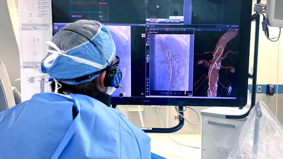
Philips Healthcare announced today it is launching a limited release of a new light-based catheter guidance system for cath labs that represents a step toward radiation-free interventional imaging. The focus of the technology right now is endovascular aortic procedures.
The company said its new LumiGuide system is being made available to specialized hospitals in Europe and the United States as a next step to gather more clinical data on its performance. The next step will be making the solution available globally.
LumiGuide uses fiber optic realShape (FORS) technology, where light is reflected along an optical fiber inside a guidewire to generate 3D, high-resolution, color images of devices inside a patient’s body in real time from any angle and in multiple views. This includes the visualization of off-the-shelf catheters. Philips said the technology allows operators to know which direction their device is facing and see where they need to go. This navigation can be done without X-ray angiography or fluoroscopy. Philips has been working on this FORS technology for several years.
"I think that FORS is one of the most exciting changes that we’ve seen with imaging certainly throughout my career,” explained Andres Schanzer, MD, a vascular surgeon at UMass Memorial Medical Center. "We are seeing wires and catheters now in the foreground in front of the [X-ray] image instead of just a lot of shades of gray. Now we can just rotate a model around to look at patient anatomy in ways that are just not possible with traditional X-ray."
In late 2023, LumiGuide was used for the first time to treat patients in Maastricht University Medical Center in the Netherlands, closely followed by the University of Alabama at Birmingham. The other U.S. centers include UMass Memorial Health and Beth Israel Deaconess Medical Center. This system was developed in close collaboration with clinical partners and was made available first to major aortic centers of excellence that perform complex aortic repairs in the U.S. and Europe.
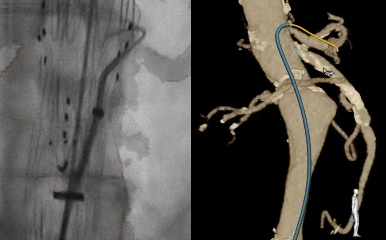
A comparison with traditional angiography, left, and the new Philips LumiGuide FORS imaging of an aorta with a stent graft.
“This technology is really transformative and is as cutting-edge as anything I’ve seen in my medical career thus far,” explained Adam Beck, MD, director of the University of Alabama at Birmingham’s Division of Vascular Surgery and Endovascular Therapy, in a statement. He has been using the FORS technology since 2021.
LumiGuide’s radiation-free technology brings potentially game-changing benefits for complex aortic procedures. In vascular surgery, physicians often perform endovascular procedures using devices like guidewires and catheters using femoral artery vascular access. For many decades, clinicians had to rely on X-ray angiography imaging systems to guide their devices through blood vessels. X-ray angiography can only produce 2D black and white images.
The bigger issue is the X-ray radiation exposure to patients, the operator and cath lab staff. Overall, digital detectors and other new technologies on angiography systems have greatly decreased radiation over the past decade. However, longer procedure times for ever more complex procedures today has caused radiation to remain as a major safety concern.
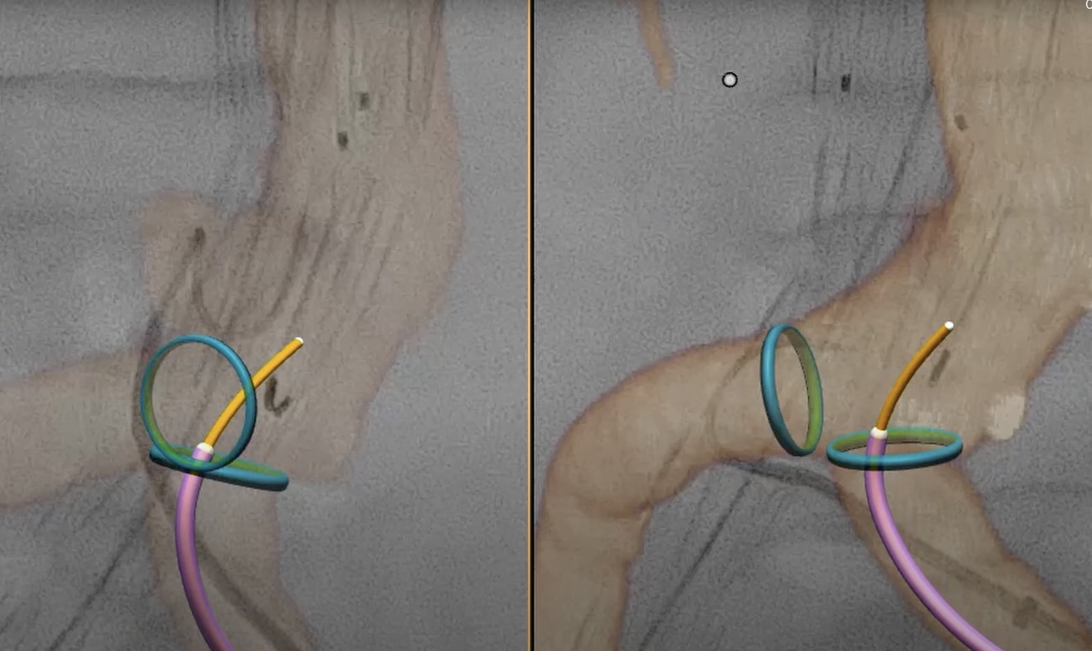
Philips LumiGuide FORS system showing 2 different views of same catheter entering the aorta from an iliac.
Users have positive things to say about the impact of FORS technology
"With LumiGuide we defiantly see more because we can see projections that were never possible before. Now the whole team, can see better what happens during the catheterization. Lumiguide helps speed up the intervention. The three-dimensional visualization of the LumiGuide catheter and wire allows easier catheterization of the target vessels," Tilo Kolbel, MD, a vascular surgeon at the German Aortic Center Hamburg, University Heart Center, explained in a video about his experience with the technolology.
Having worked on this technology for the past several years, Philips has been able show promising study results around the world. Following a limited release to nine aortic centers, more than 900 patients have undergone procedures. One site has conducted a historic cohort comparison, showing a 37% reduction in complex aortic procedure time, and a 56% reduction in radiation exposure (DAP) compared to X-ray.[1]
"If we can see more, we can proceed more quickly and more confidently. In effect, LumiGuide is a 3D human GPS system powered by light,” Atul Gupta, MD, chief medical officer for image guided therapy and precision diagnosis at Philips, and practicing interventional radiologist, said in a statement.
Integrating AI to enable automatic FORS registration of catheters
LumiGuide works exclusively with compatible Philips interventional systems such as Azurion and is the second-generation solution to use FORS. The first-generation system was used at nine centers to building data and clinical feedback. Based on that, LumiGuide incorporates new time-saving features such as eliminating the need for physicians to manually register their devices with their image-guided therapy platform. Instead, LumiGuide uses AI-based recognition to register the guidewire quickly and efficiently, helping to increase accuracy while speeding up procedure time.
“This AI-based semi-automatic registration is very quick and accurate, even in the presence of stent grafts. Especially if there is a need to re-register the device being guided in the patient’s body during the procedure, it is extremely helpful,” explained Geert Willem Schurink, a vascular surgeon at Maastricht University Medical Center, who performed the first surgical procedure with LumiGuide.

Dave Fornell has covered healthcare for more than 17 years, with a focus in cardiology and radiology. Fornell is a 5-time winner of a Jesse H. Neal Award, the most prestigious editorial honors in the field of specialized journalism. The wins included best technical content, best use of social media and best COVID-19 coverage. Fornell was also a three-time Neal finalist for best range of work by a single author. He produces more than 100 editorial videos each year, most of them interviews with key opinion leaders in medicine. He also writes technical articles, covers key trends, conducts video hospital site visits, and is very involved with social media. E-mail: [email protected]