
PHOTO GALLERY: The trends and technologies at ASE 2023
This is a visual overview of some of the highlights and new technology featured at the American Society of Echocardiography (ASE) 2023 meeting, which took place June 23-26 in National Harbor, Maryland. The show's biggest trends included the use of artificial intelligence (AI) to enhance imaging and speed workflow, increased interest in diagnosing hypertrophic cardiomyopathy (HCM) and amyloid and advances in 3D and structural heart imaging.
Read the captions for more details.
ASE is the largest dedicated cardiac ultrasound conference. It featured 2,034 attendees this year, with numbers starting to return to normal after the pandemic. Attendance was 2,507 in 2019. This recovery to near pre-pandemic numbers has been seen across medical conferences.
Deployed Watchman left atrial appendage (LAA) occluder visualized on Siemens' 3D intracardiac echo (ICE) technology demonstrated on the expo floor at ASE 2023. ICE is being used more and more to help perform LAA occluder, mitral and tricuspid repair procedures now that its 3D image resolution has greatly improved in the past few years.
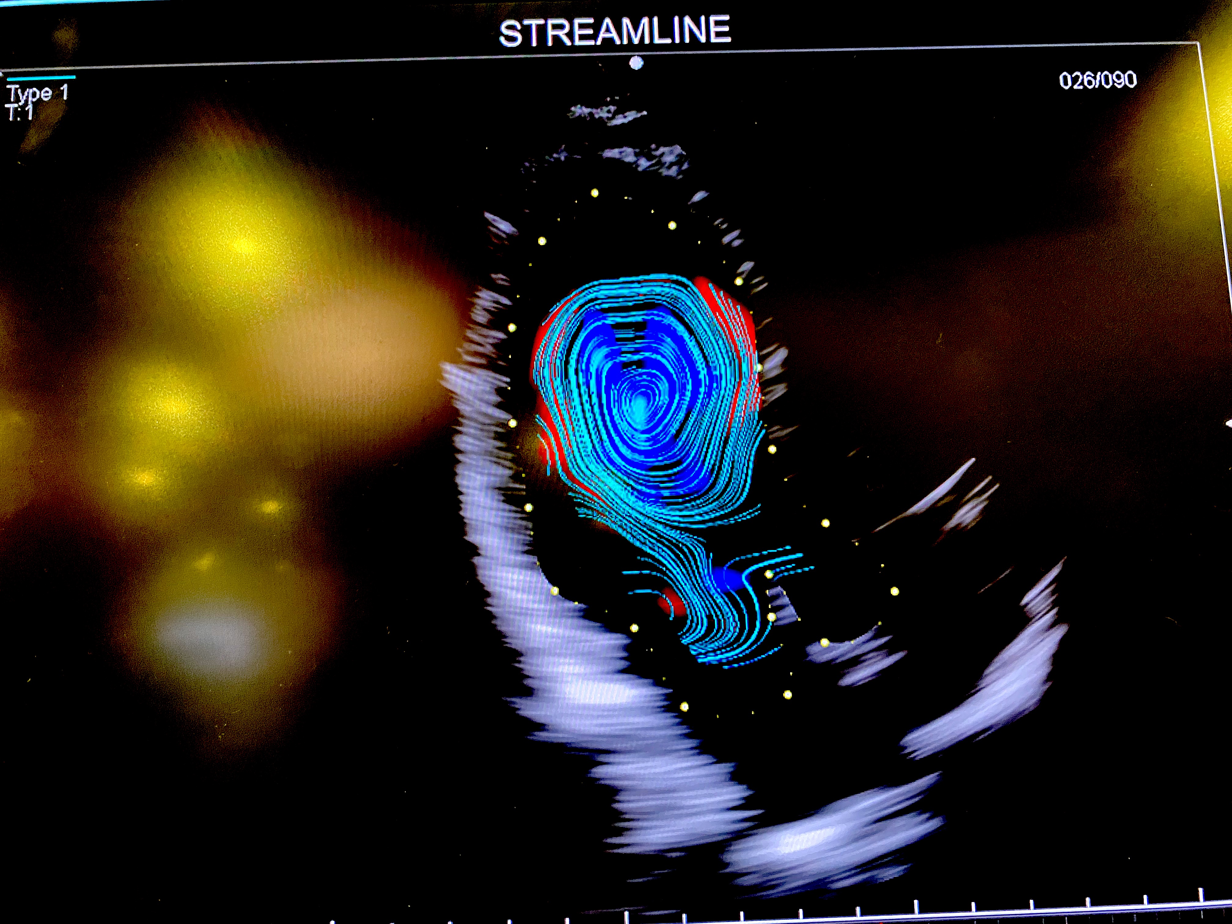
A vortex of blood flow inside the left ventricle visualized using the Streamline view of vector flow imaging on the Fujifilm Arietta 750 Cardiovascular ultrasound system. Several vendors now have similar ultrasound blood flow speckle tracking technologies that are primarily being used in research to understand the impact of vortices and sheer stress on the heart caused by various diseases. It is believed by many cardiology experts that this type of visualization may be able to detect disease or progression of disease long before a patient becomes symptomatic and one day help better risk stratify patients.
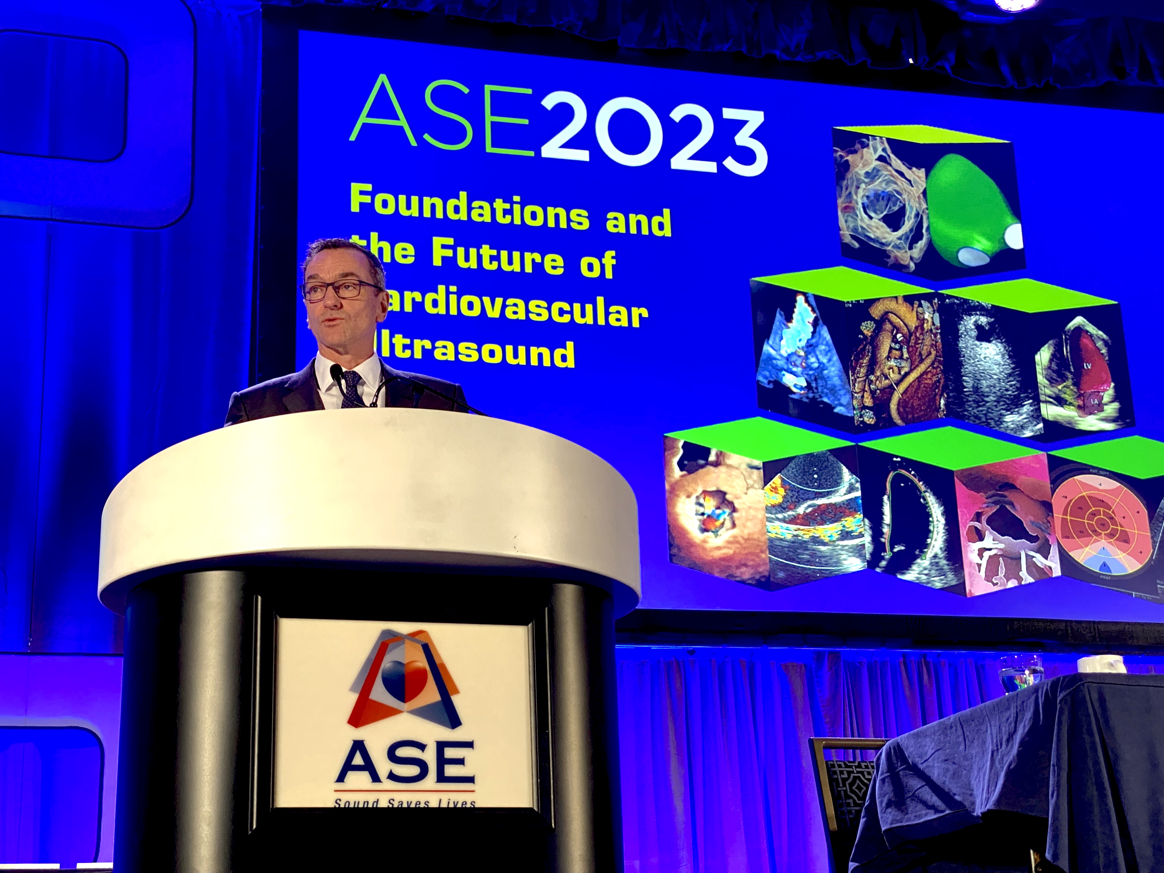
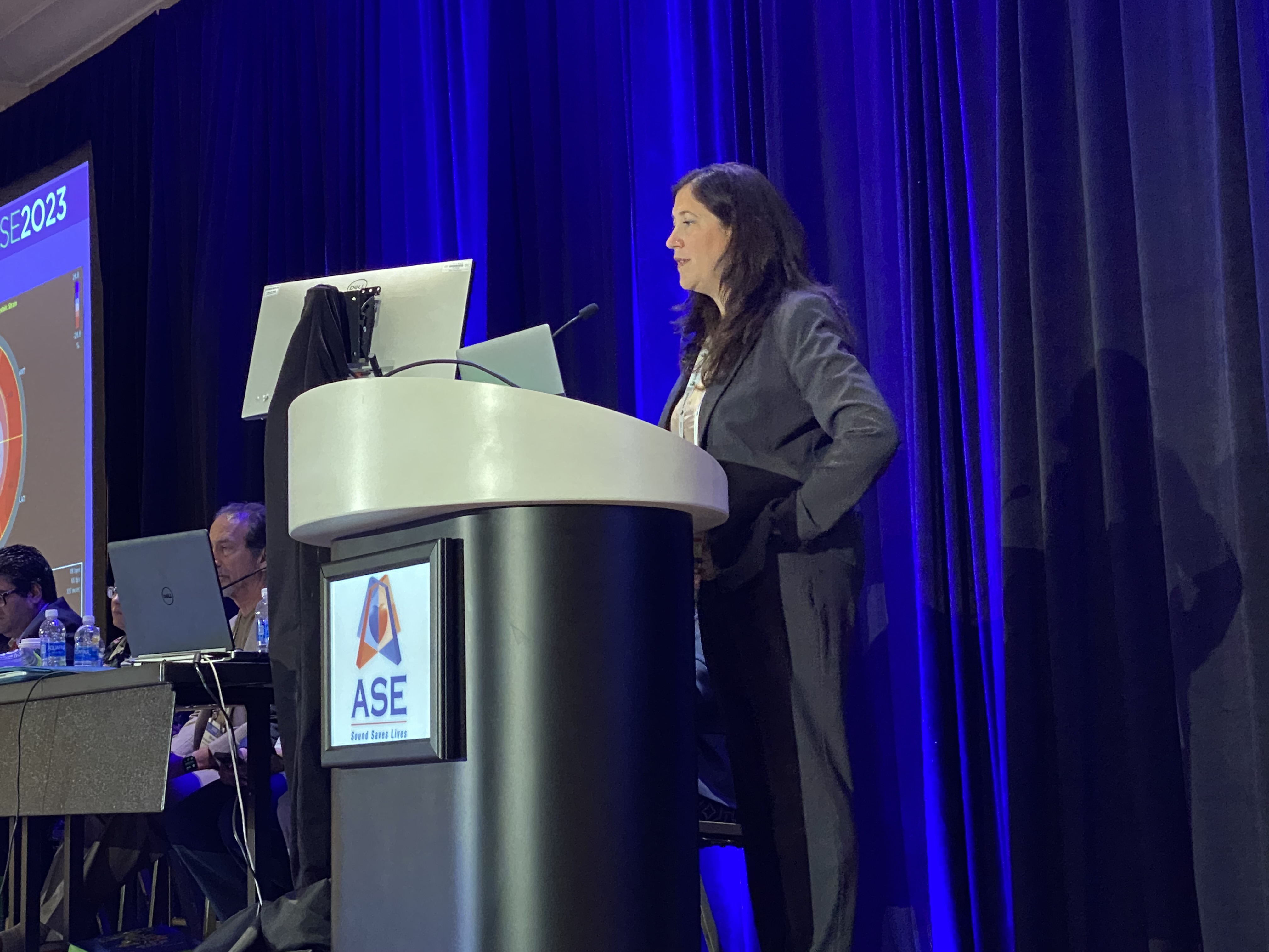
ASE President Stephen Little, MD, of Houston Methodist delivers the State of ASE opening session. Highlights included a call for echocardiography to embrace new technologies such as AI and telemedicine to help speed workflows to see more patients. Little said echo is facing growing shortages of sonographers while at the same time facing growing patient volumes. ASE is now partnering with numerous vendors to push new technologies forward and created task forces to concentrate on recommendations for AI, ICE and other areas.
A simulated Edwards Sapien TAVR valve in the mitral position in a 3D echo image being used for transcatheter mitral valve replacement (TMVR) planning from Pie Medical. The software uses echocardiograms for sizing the valve and to determine if the valve will block the patient's left ventricular outflow track. It can help heart teams decide if a patient is a candidate for TMVR or if additional procedures like SESAME or LAMPOON are needed.
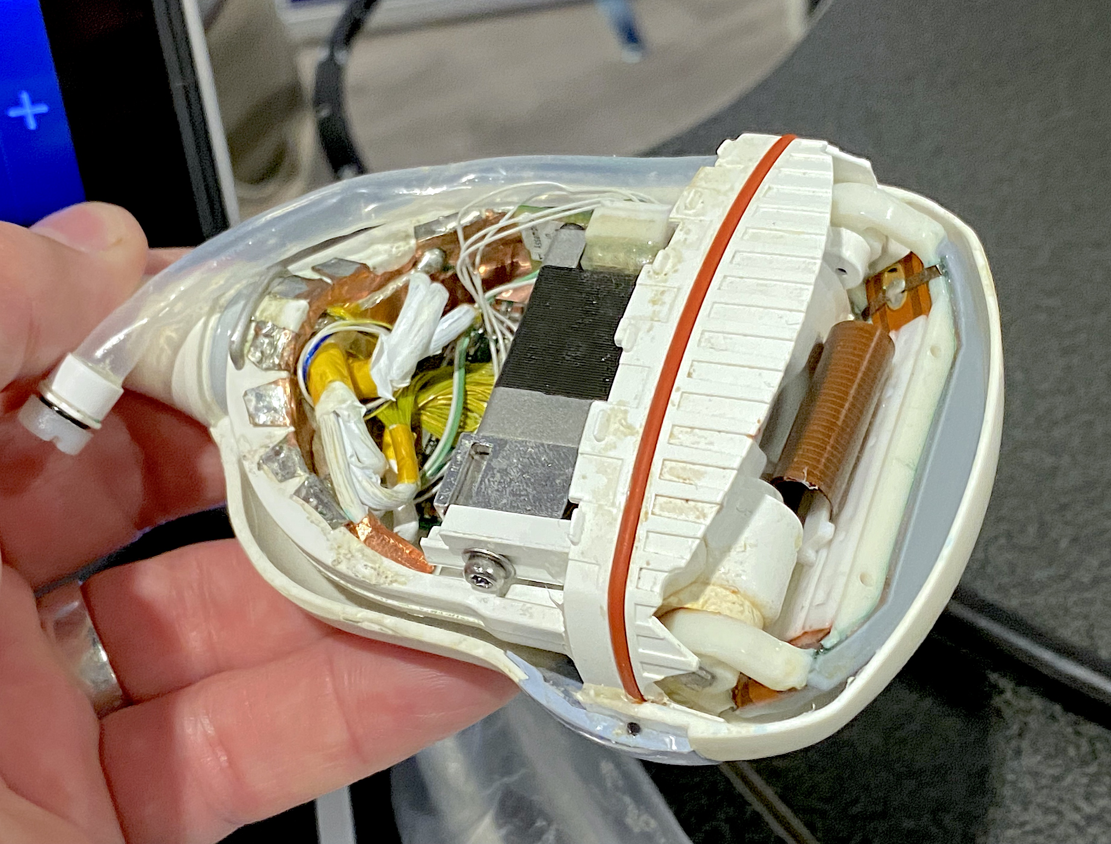
A cutaway view of an ultrasound transducer displayed by the repair service Probo on the ASE expo floor.
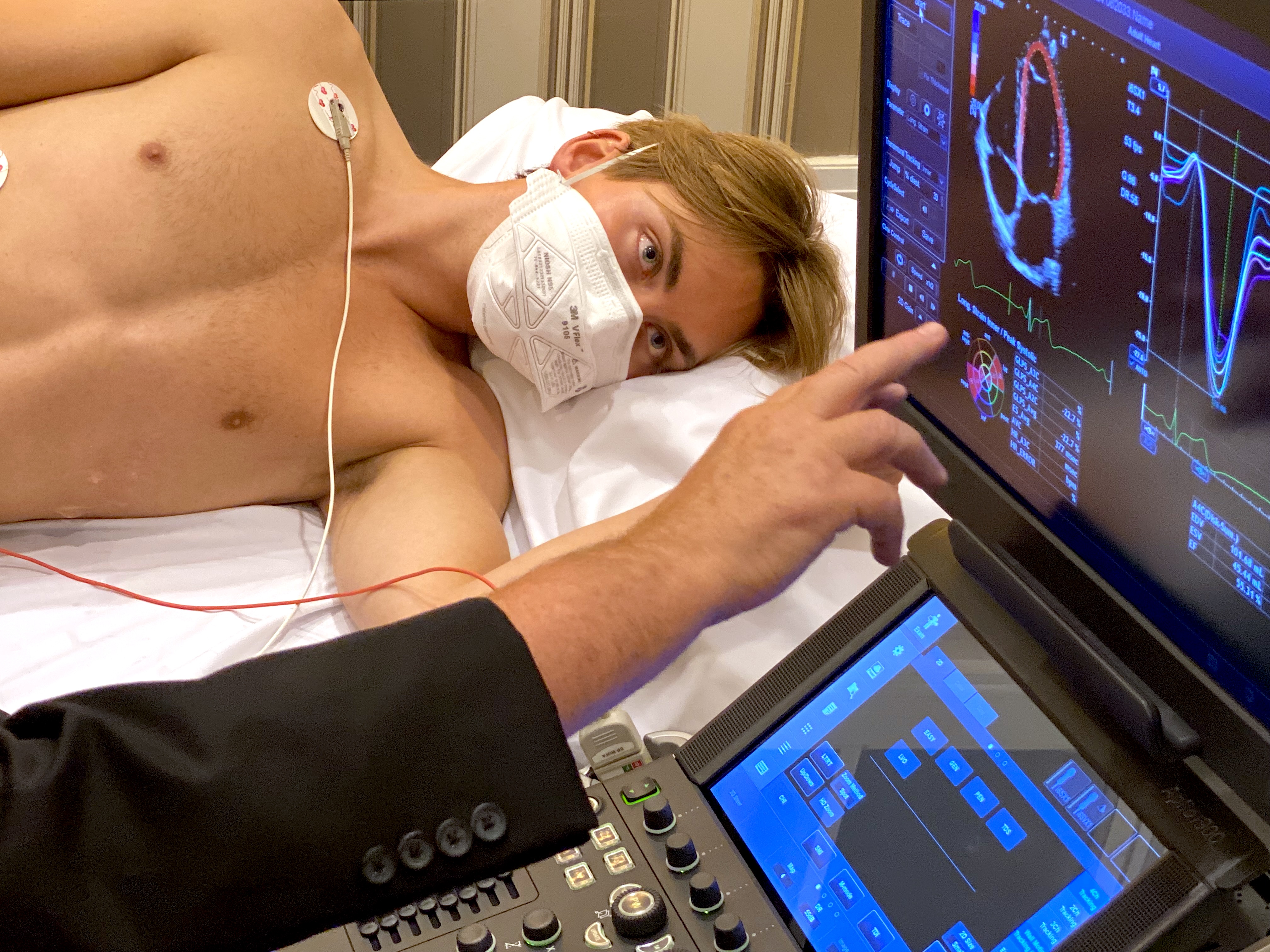
A cardiac strain exam being explained during one of several hands-on training courses hosted by ASE.

Sunset over the Potomac River in National Harbor, Maryland, where ASE 2023 was held.
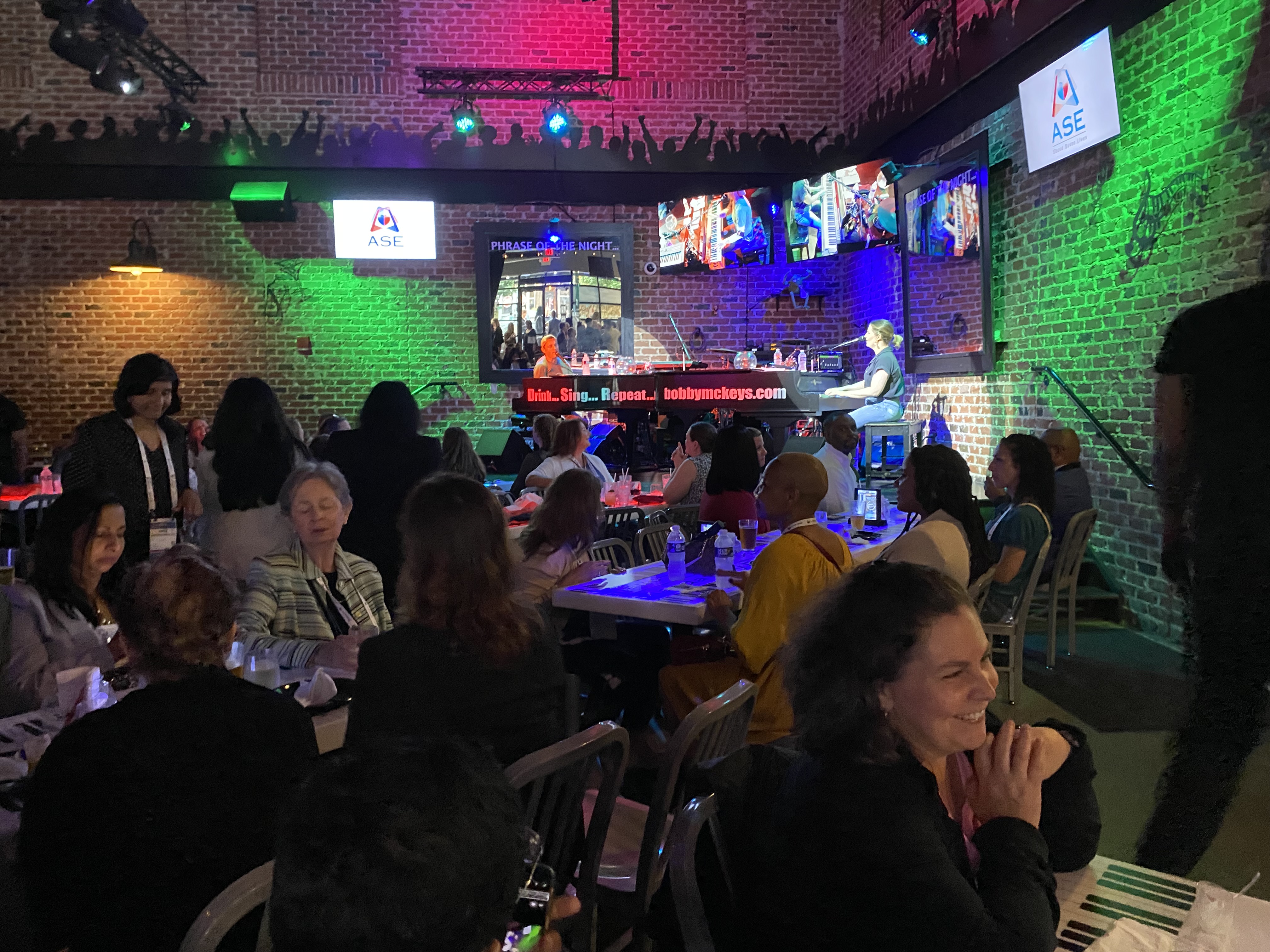
ASE 2023 welcome reception and social event at Bobby McKey's Dueling Piano Bar in National Harbor on the first night of the conference. So many attendees came to the event that they had a waiting line out front for part of the evening.
A patient being scanned during a live, hands-on strain echo sonographer training session at ASE 2023.
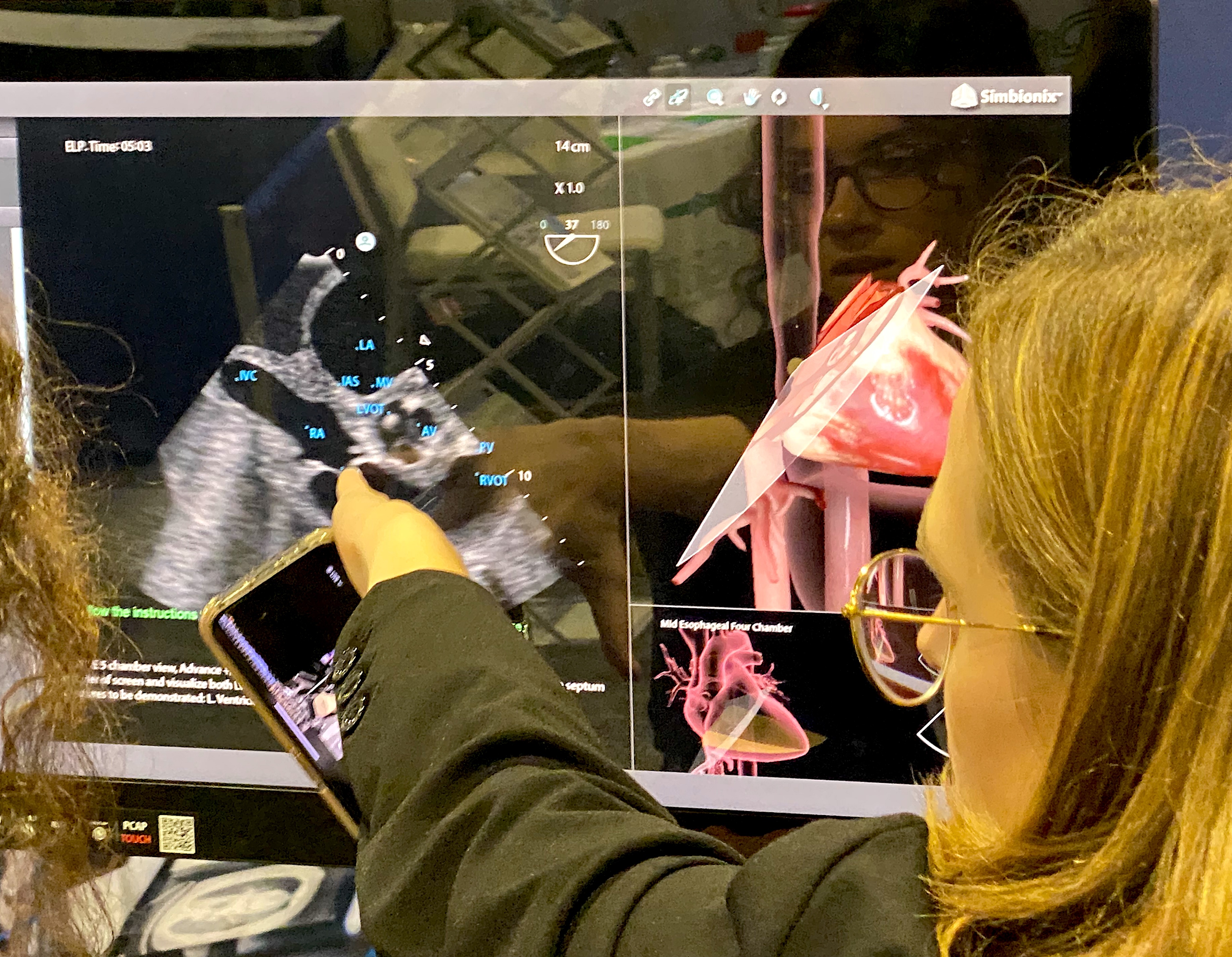
A TEE simulator for sonographer training shown by SurgicalScience on the ASE expo floor.
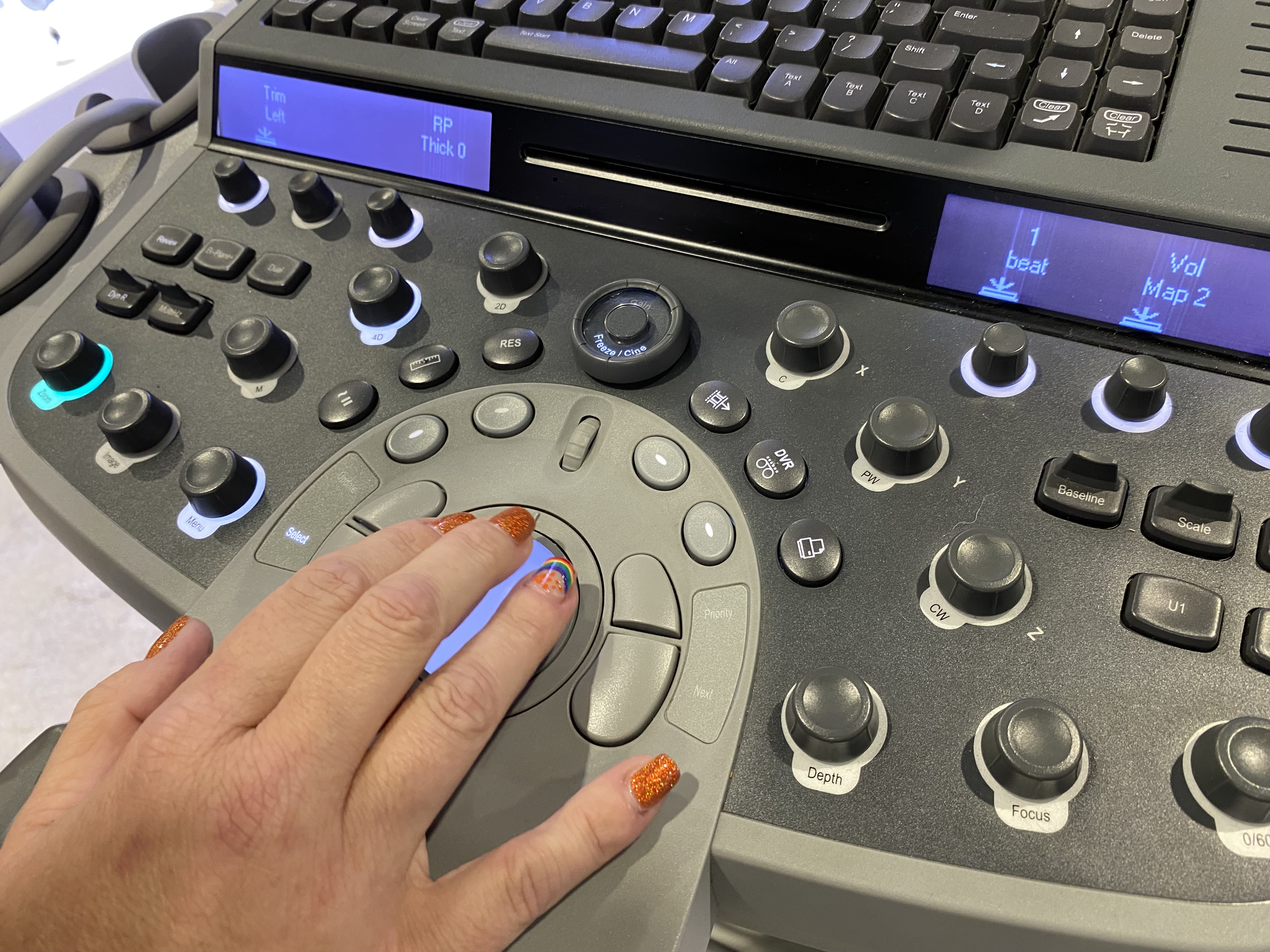
A sonographer uses the trackball on a Siemens ultrasound system during a booth demonstration on the expo floor.
Ritu Thamman, MD, leads a Women in Echo session on how to address bullying issues in the workplace. During the session, numerous audience members shared stories from their echo labs about problems they or coworkers experienced and what they did to report and document the issues, including how some prepared legal cases.
The Philips VeriSight 3D intracardiac echo (ICE) probe. 3D ICE has gained a lot of attention the past couple years because the new technology offers the ability to visualize procedures inside the heart with image quality almost as good as TEE. ICE is seeing use in LAA occluder procedures and mitral and tricuspid valve repairs.
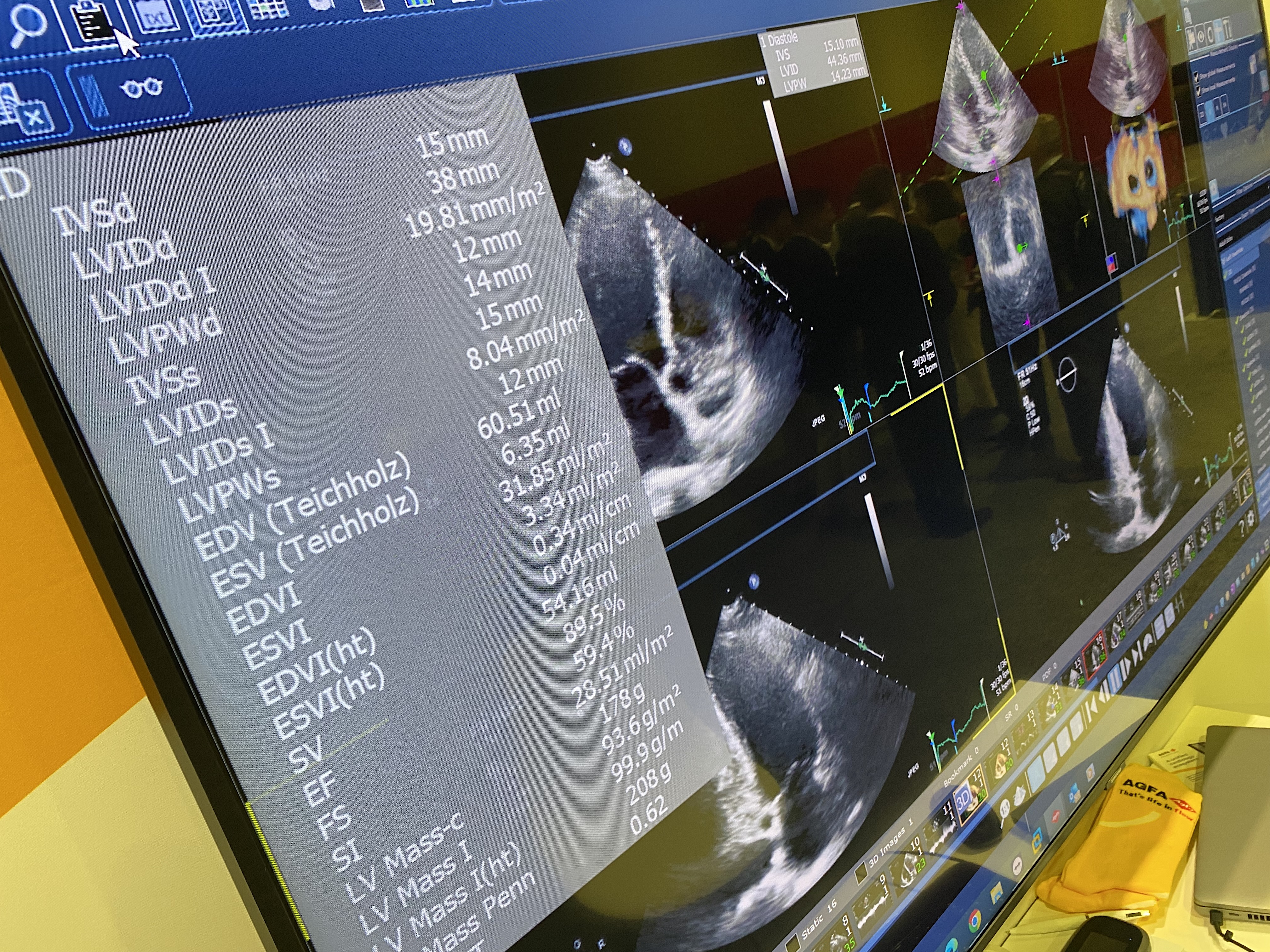
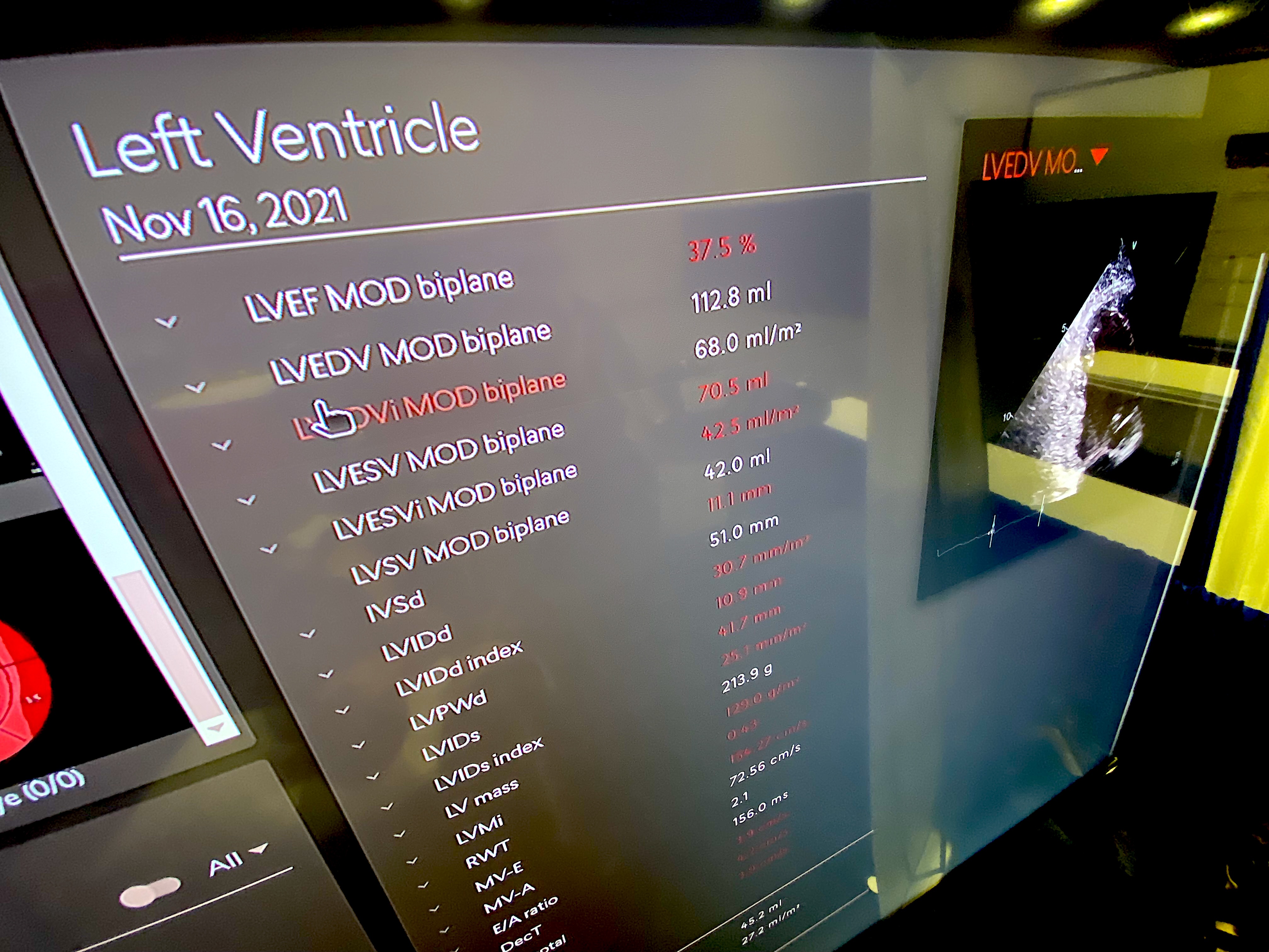
Automated echo measurements on the Agfa echo reporting system in its enterprise cardiovascular information system (CVIS) demonstrated at ASE 2023. These types of automation were highlighted across the expo floor to help improve accuracy, consistency and speed workflows.
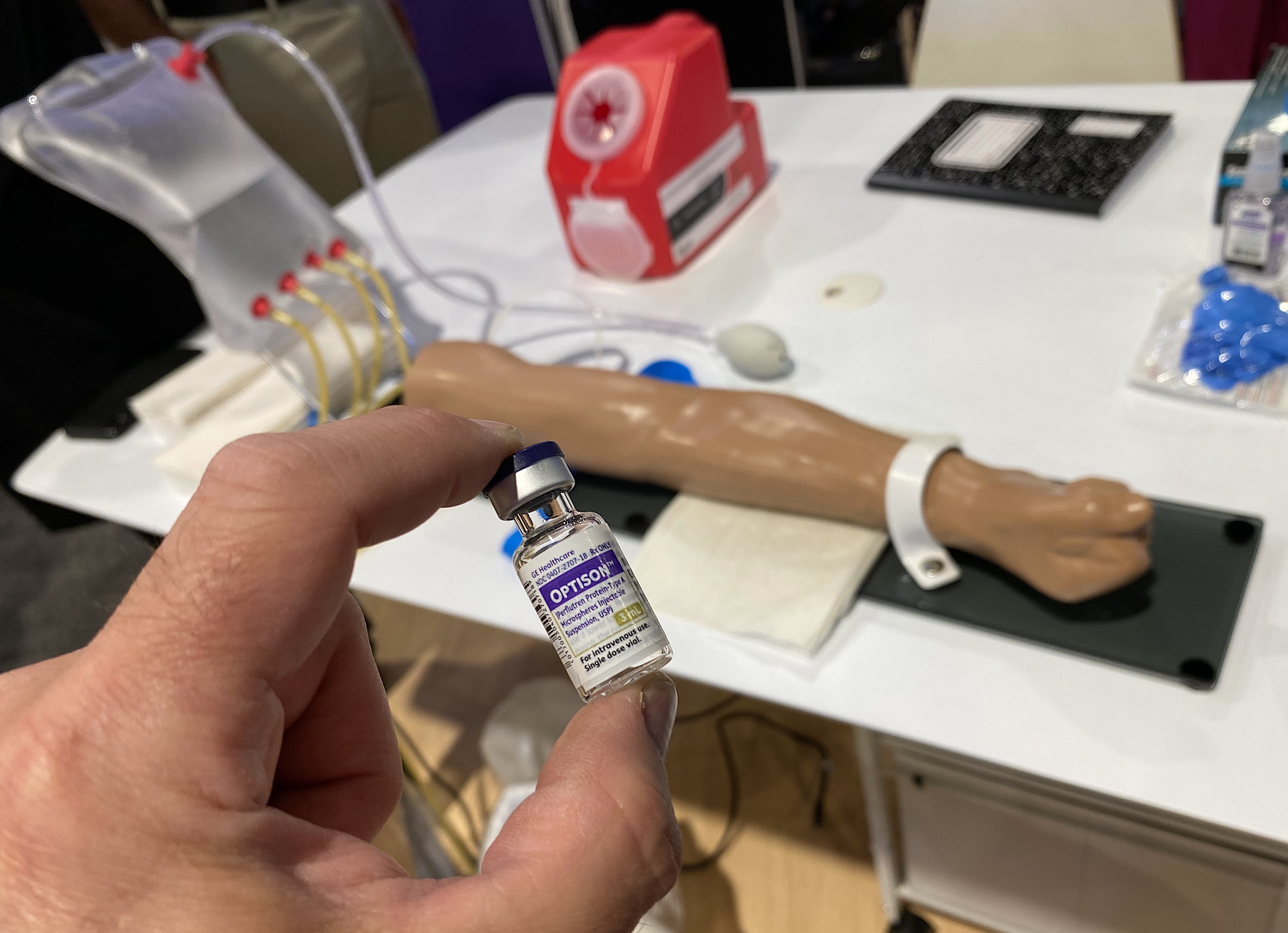
Optison cardiac ultrasound bubble contrast image enhancing agent and an IV insertion training dummy in the GE booth at ASE 2023. Echo contrast can help improve the diagnostic quality of cardiac ultrasound, especially in obese patients, but its access is limited when nurses are required to insert the IV line for the contrast. ASE and vendors want to see expanded training to enable sonographers to insert the IV lines themselves.
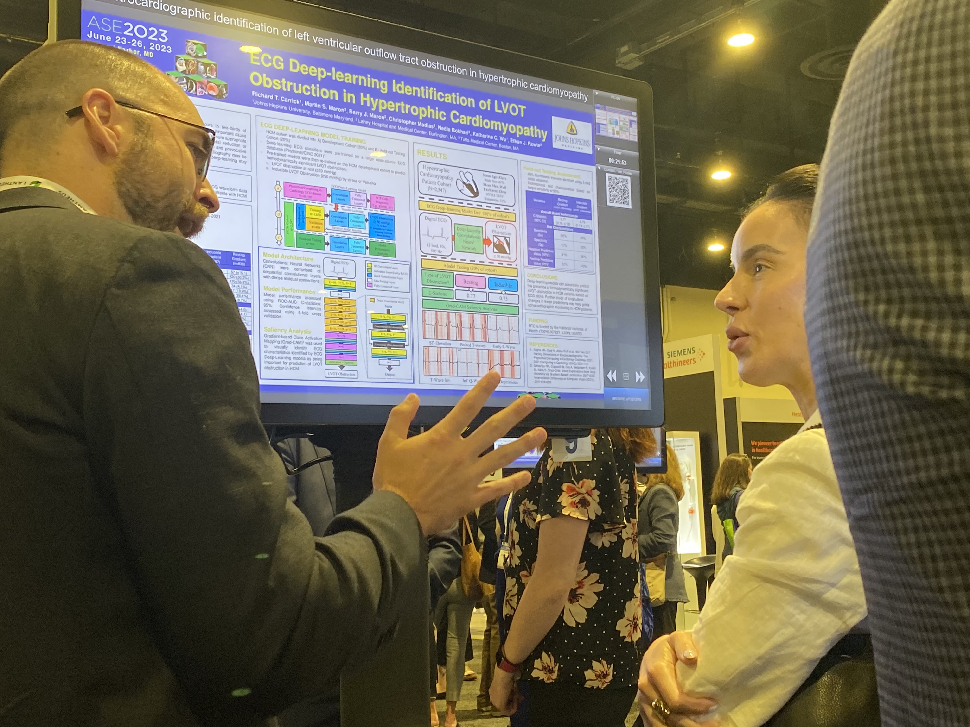
ASE attendee listens as a researcher explains their poster on the expo floor at ASE 2023.
An example of AI guidance technology to help novice ultrasound users perform a diagnostic quality echo on the Echonous Kosmos POCUS system. The green bars show the quality of the imaging window and anatomy to capture imaging of the heart is specific views. The system also has a thumbnail image of the chest and transducer to show how to move it and get the correct views.
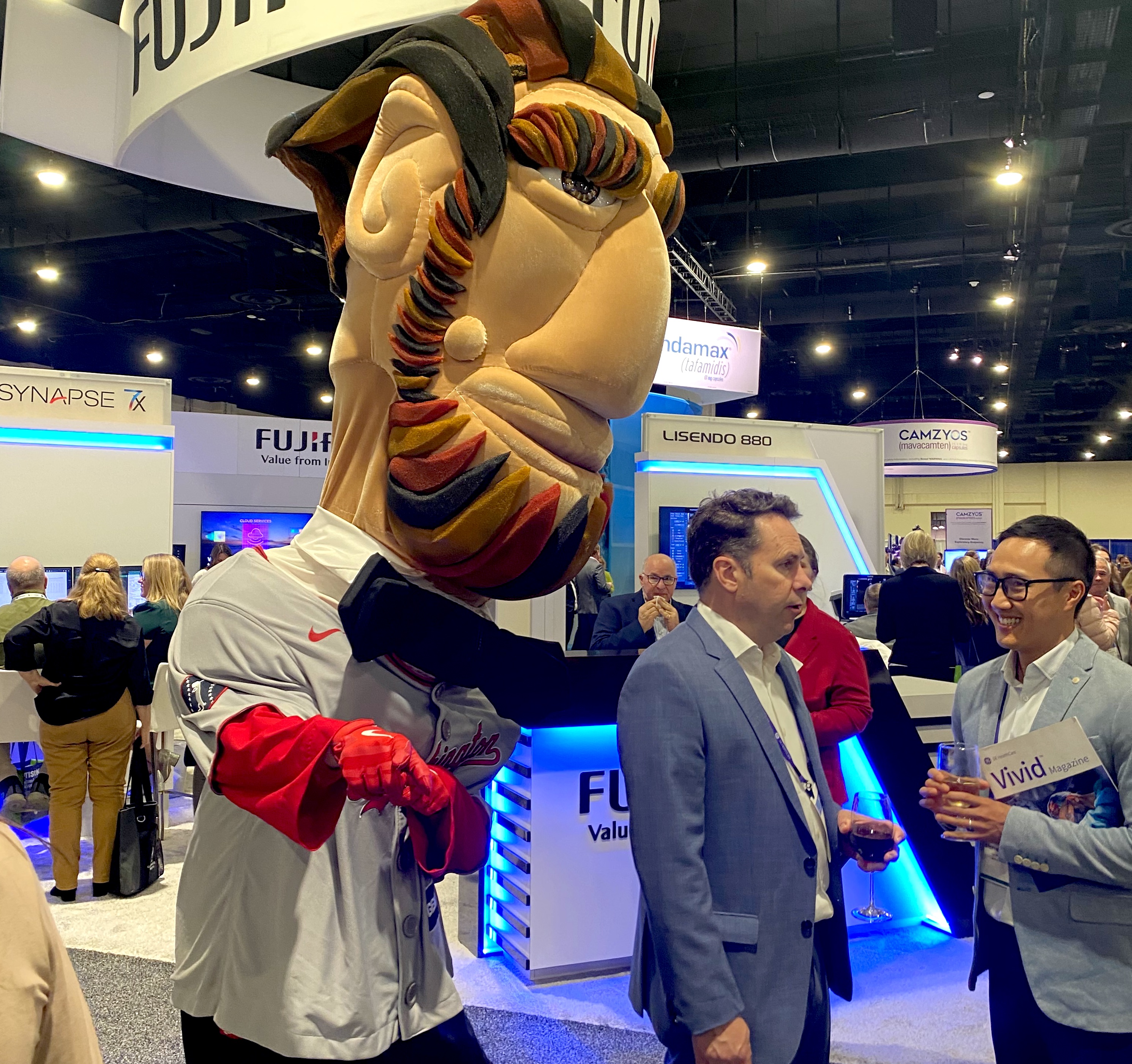
The mascot of the Washington Nationals, Abe Lincoln, causing antics and getting selfies with ASE attendees on the expo floor.
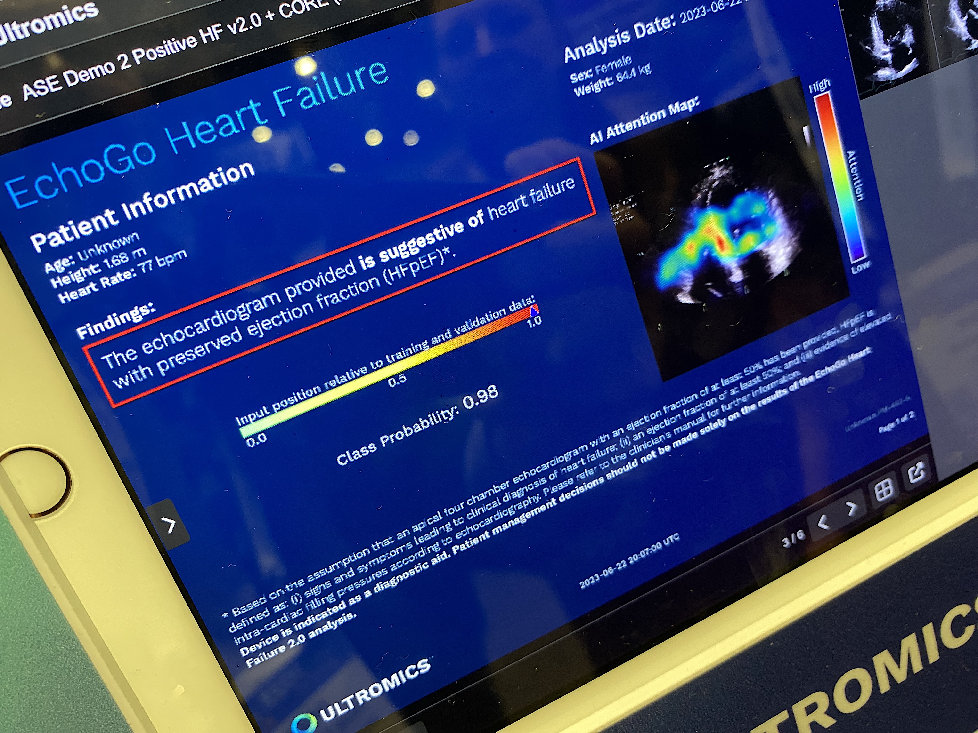
The Ultromics EchoGo Heart Failure AI algorithm to automatically detect if a patient has heart failure from an echocardiogram. The colored areas of the image are a heat map showing the areas of interest on the cardiac ultrasound image the AI used to make the suggested diagnosis.
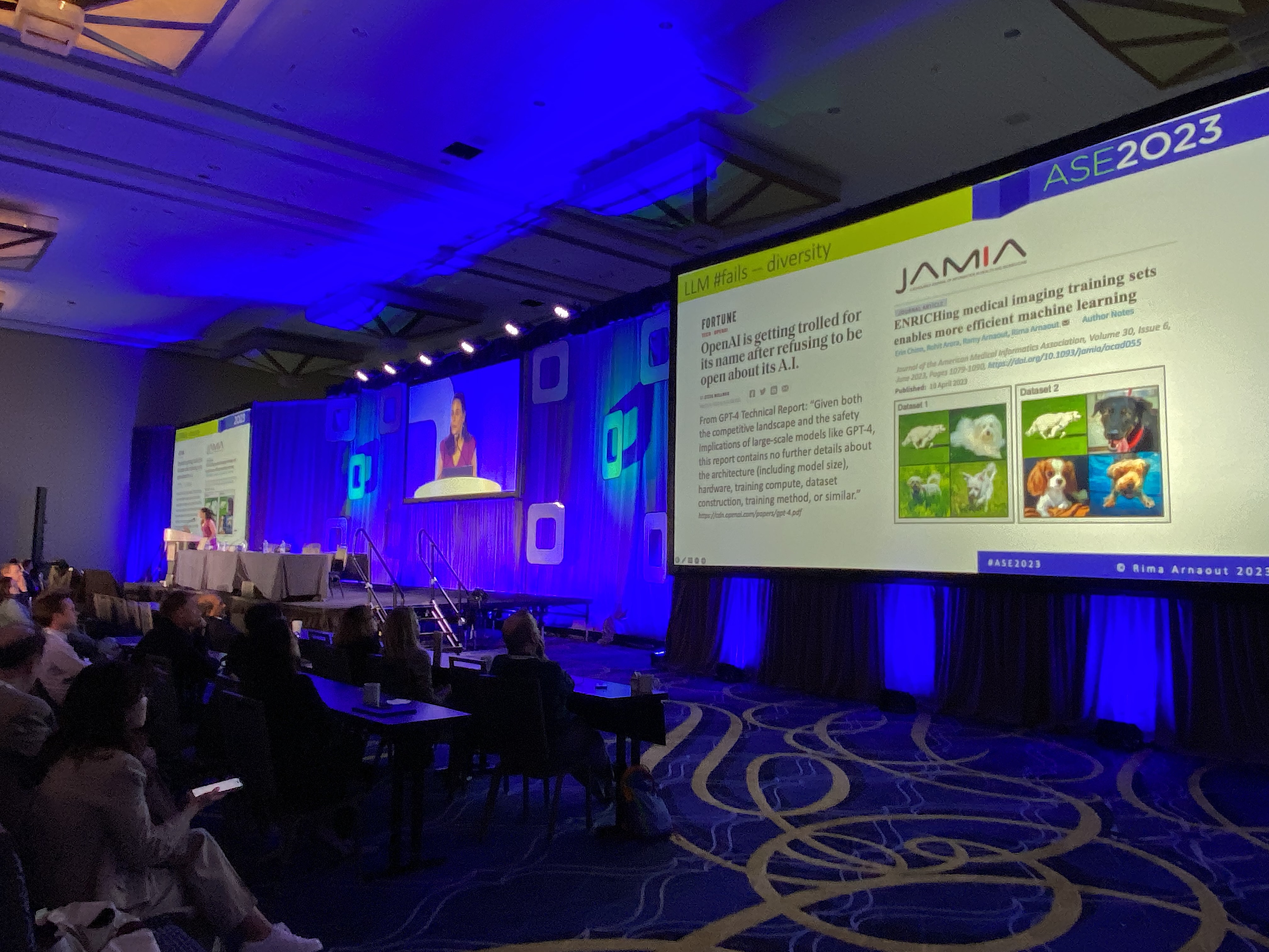
The ASE main presentation theater during a session on how artificial intelligence is impacting cardiac ultrasound.
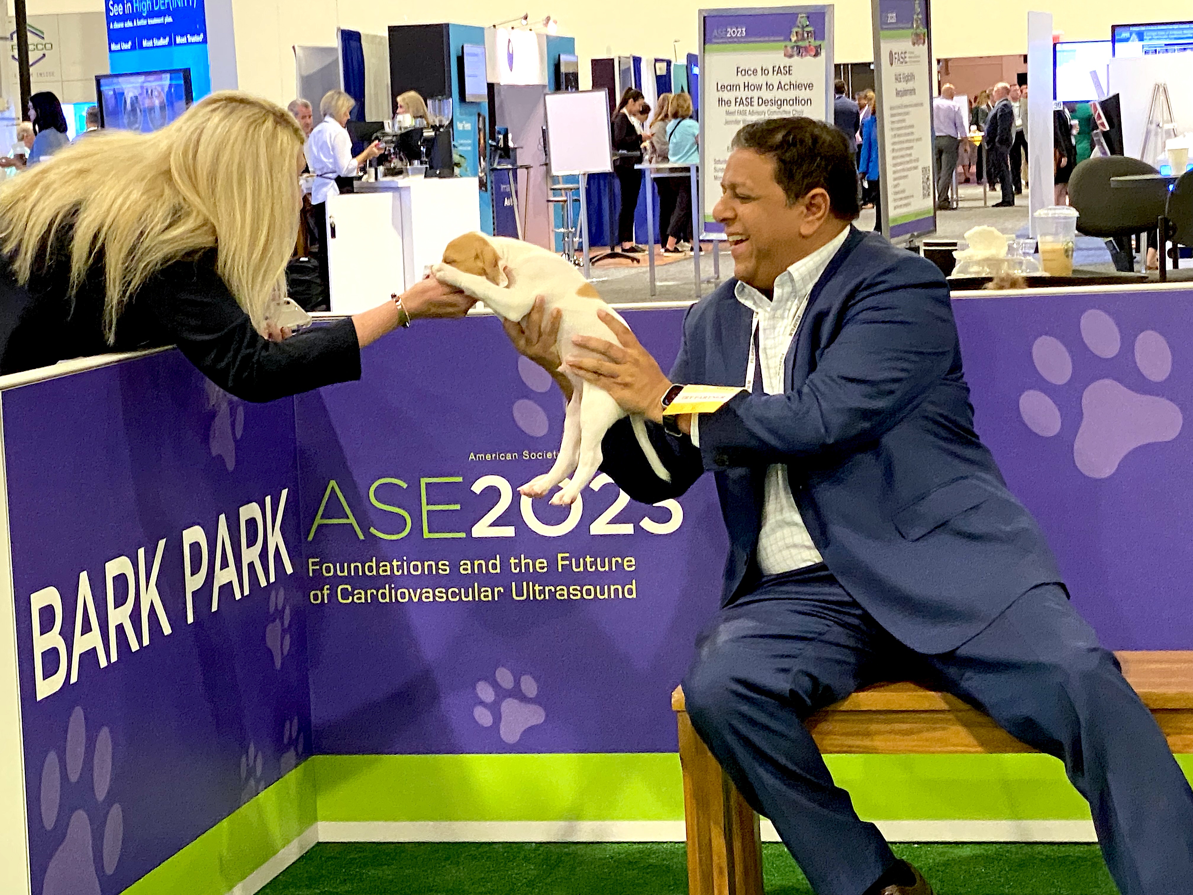
ASE 2023 attendees play with puppies at the Bark Park on the expo floor.
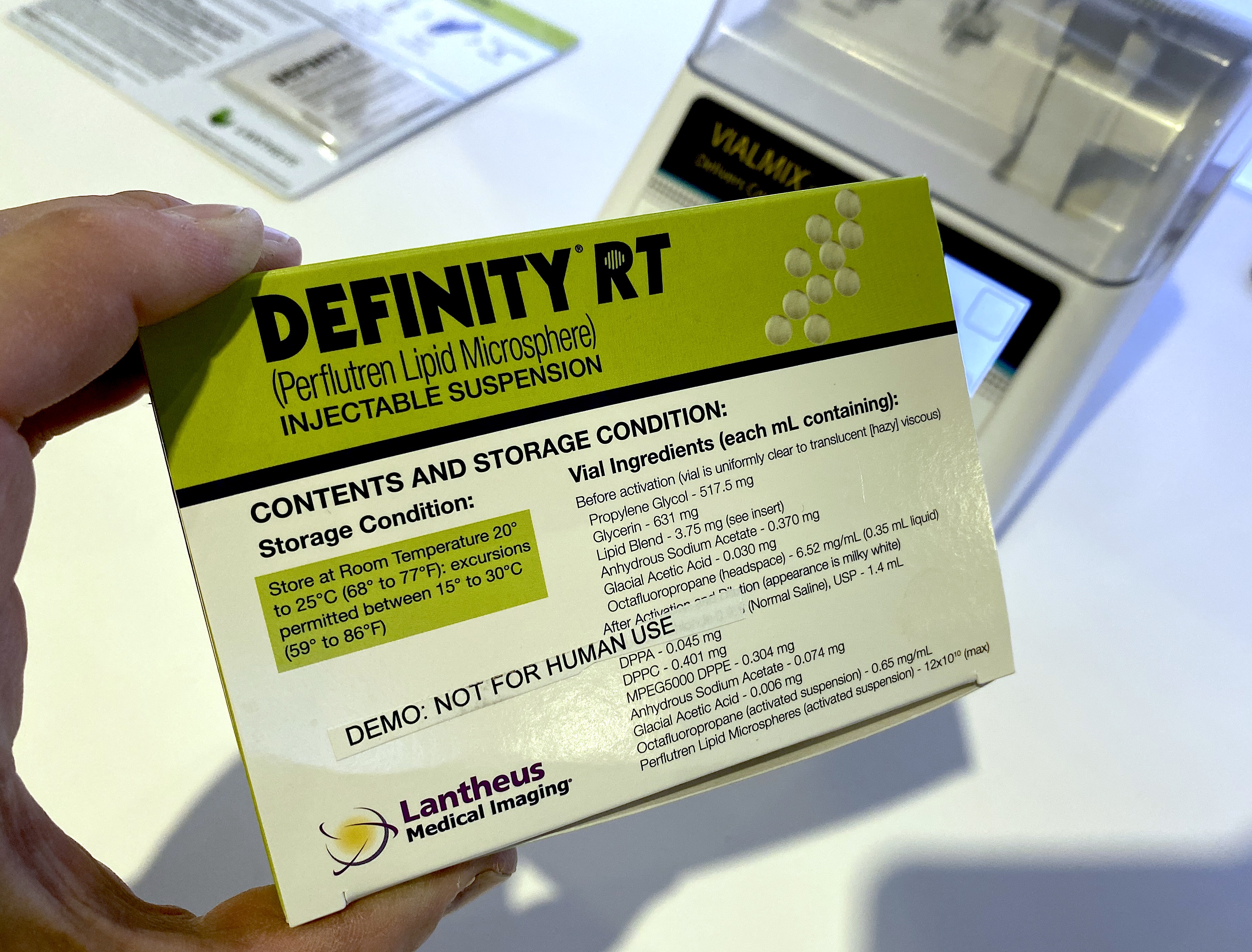
Definity echo bubble contrast in the Lantheus booth on the ASE expo floor.
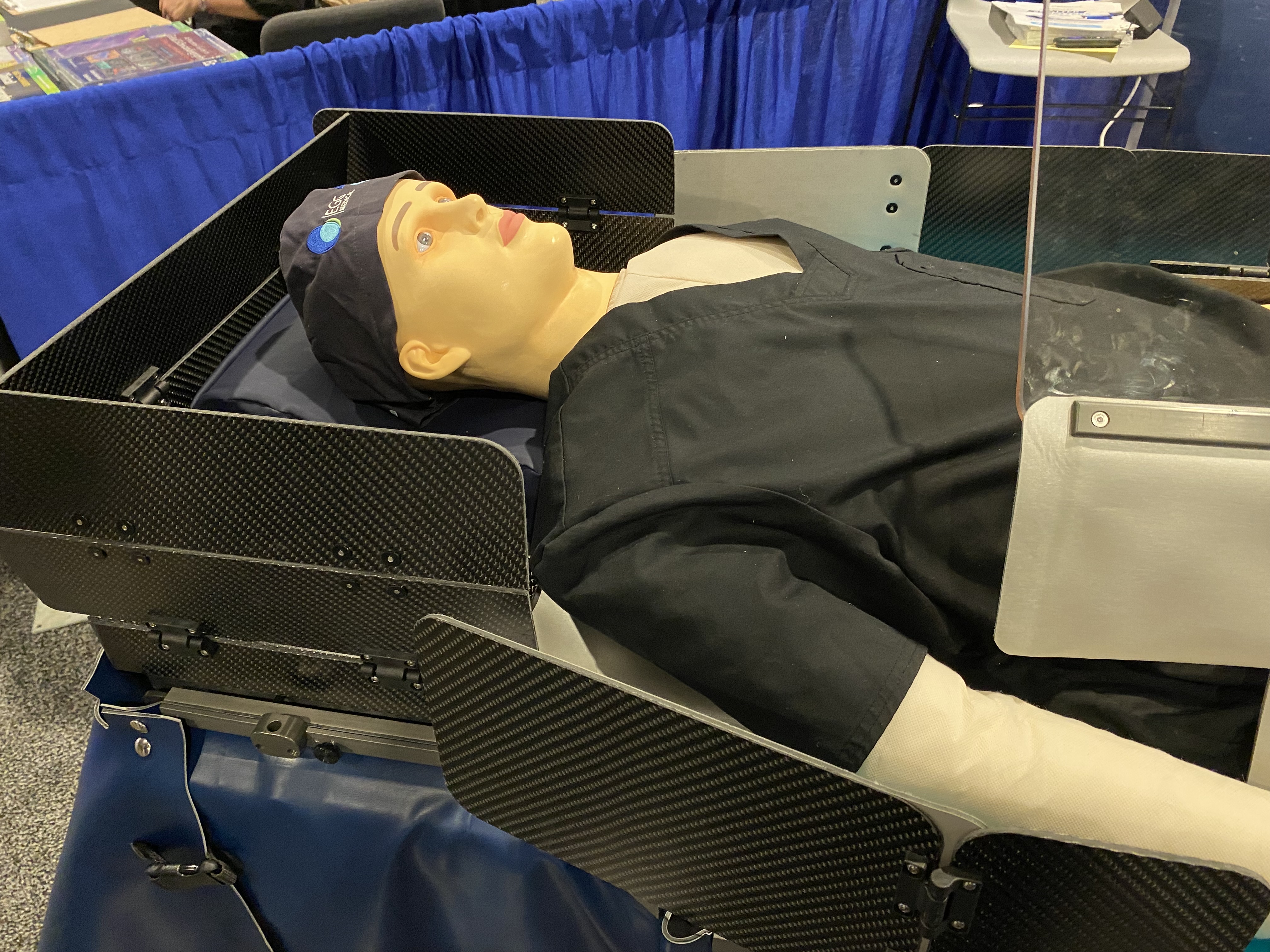
Radiation protection for the cath lab to protect the echocardiographer during structural heart procedures. Interventional echocardiographers are exposed to more radiation during procedures than the interventional cardiologist because of the position they sit at next to and often over the patient to guide the TEE probe. This bed-mounted system of shields is made of lightweight carbon fiber and bismuth. Egg Medical showed this in their booth on the expo floor.
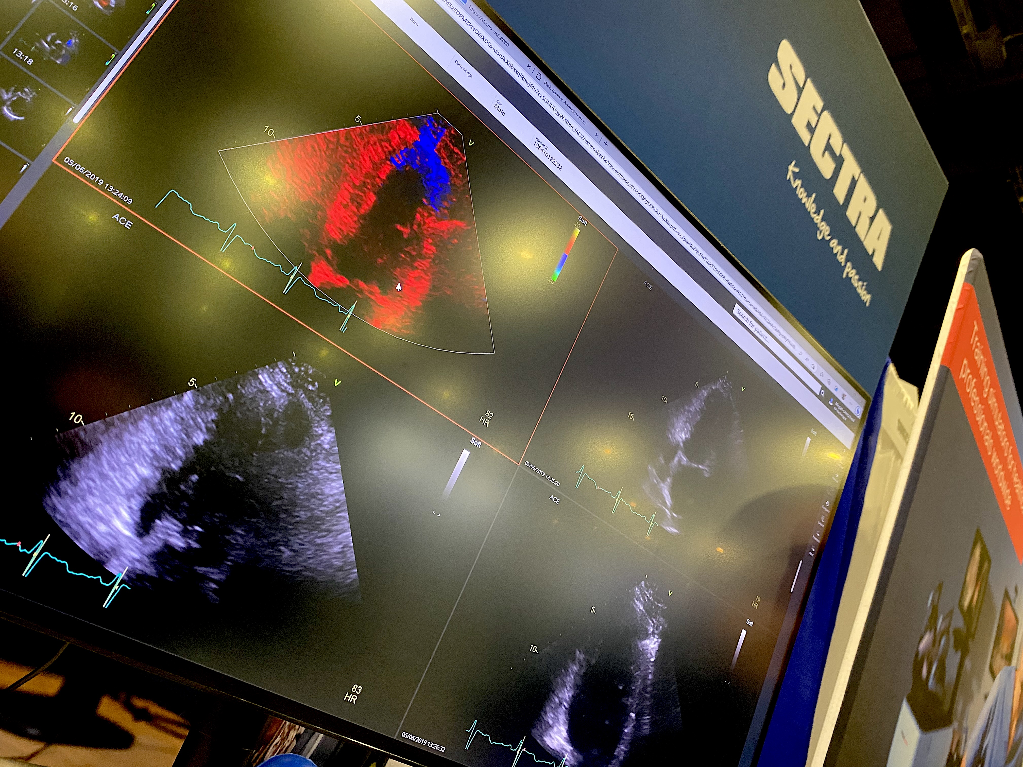
Cardiovascular information system (CVIS) and enterprise imaging IT vendor Sectra showed how its echo reporting system can help save time with workflow efficiencies.
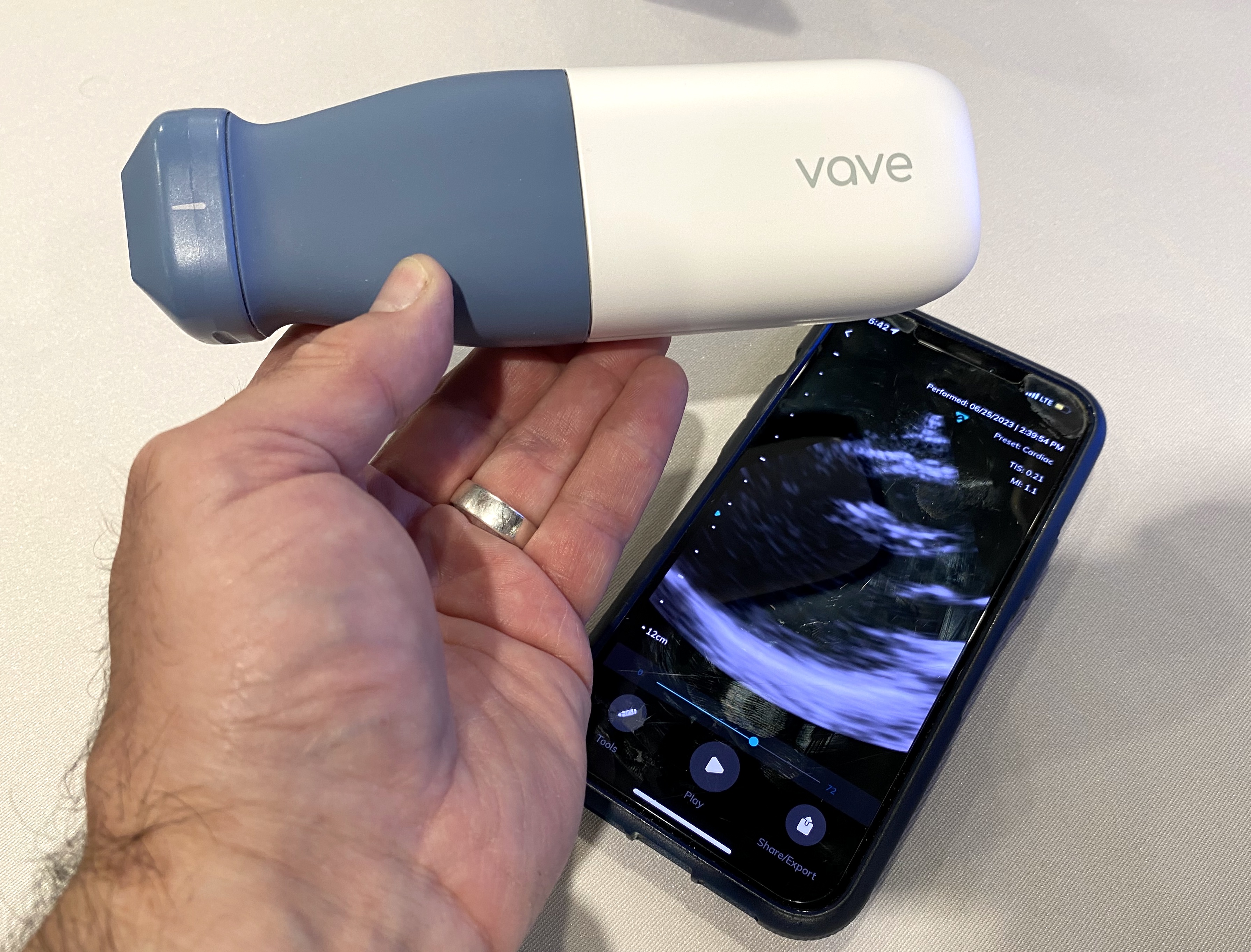
The Vave wireless point of care ultrasound (POCUS) transducer system that works with an app to turn a smartphone into a portable ultrasound system. Four of these types of systems were demonstrated on the ASE expo floor. They are helping to propel the growing use of POCUS to perform quick cardiac exams and triage outside of echo labs.
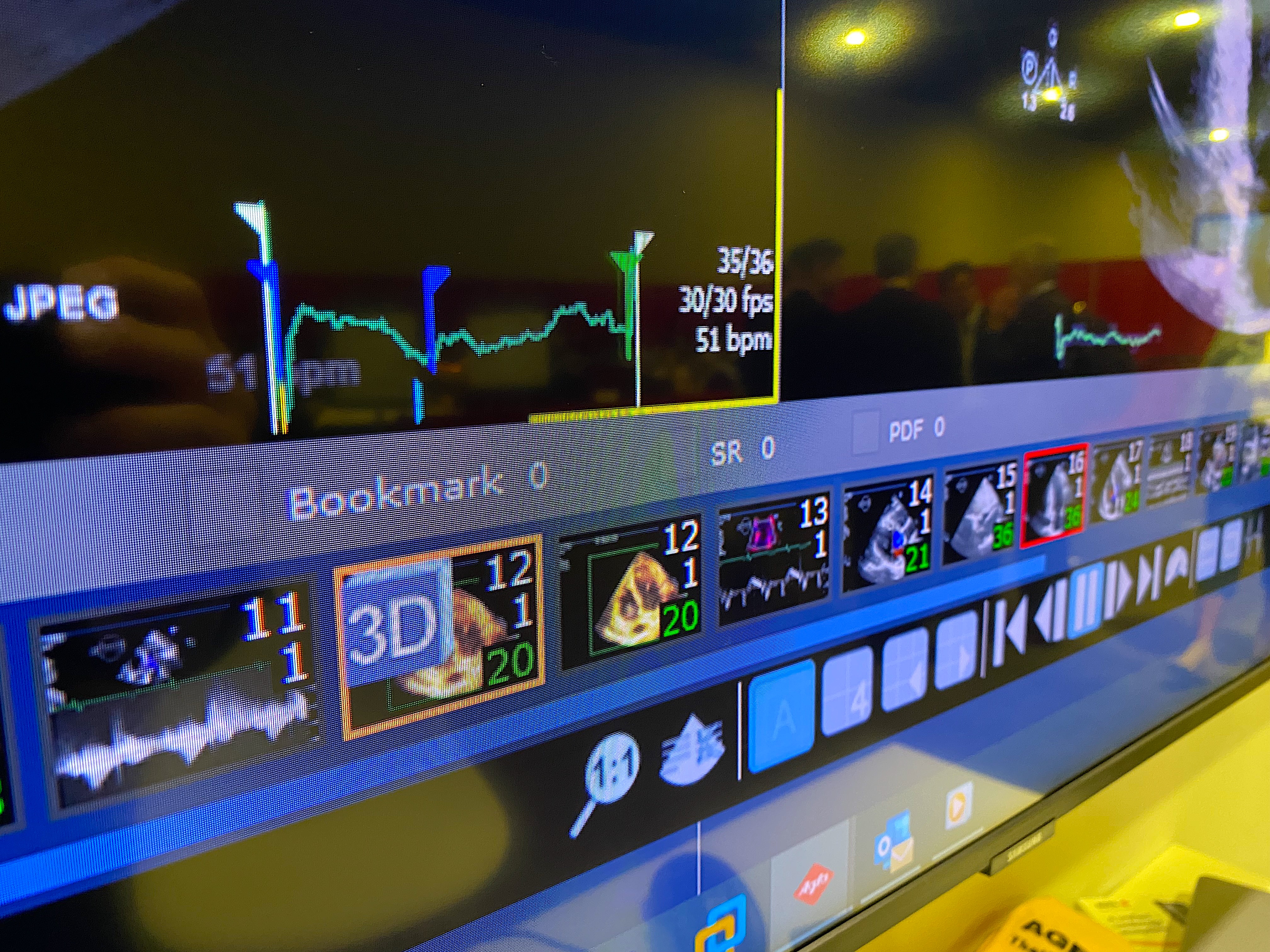
Patient thumbnail view timeline of prior exams for easy access under a current cardiac ultrasound exam on the Agfa cardiovascular information system (CVIS) echo reporting system.
Ted Abraham, MD, co-director of the UCSF HCM Center of Excellence and director of the adult echo lab, shares ideas on next steps to improve HCM care. Interest to increase screening and treatment of HCM has exploded in the past year since the FDA cleared the first drug to treat the disease. During the ASE-sponsored HCM Forum, experts in imaging and therapy shared concerns and possible solutions to increase training to perform screenings, risk stratification, roles for multimodality imaging, procedural planning the need for standardization for reporting and invasive treatments and the desire to create an HCM registry.
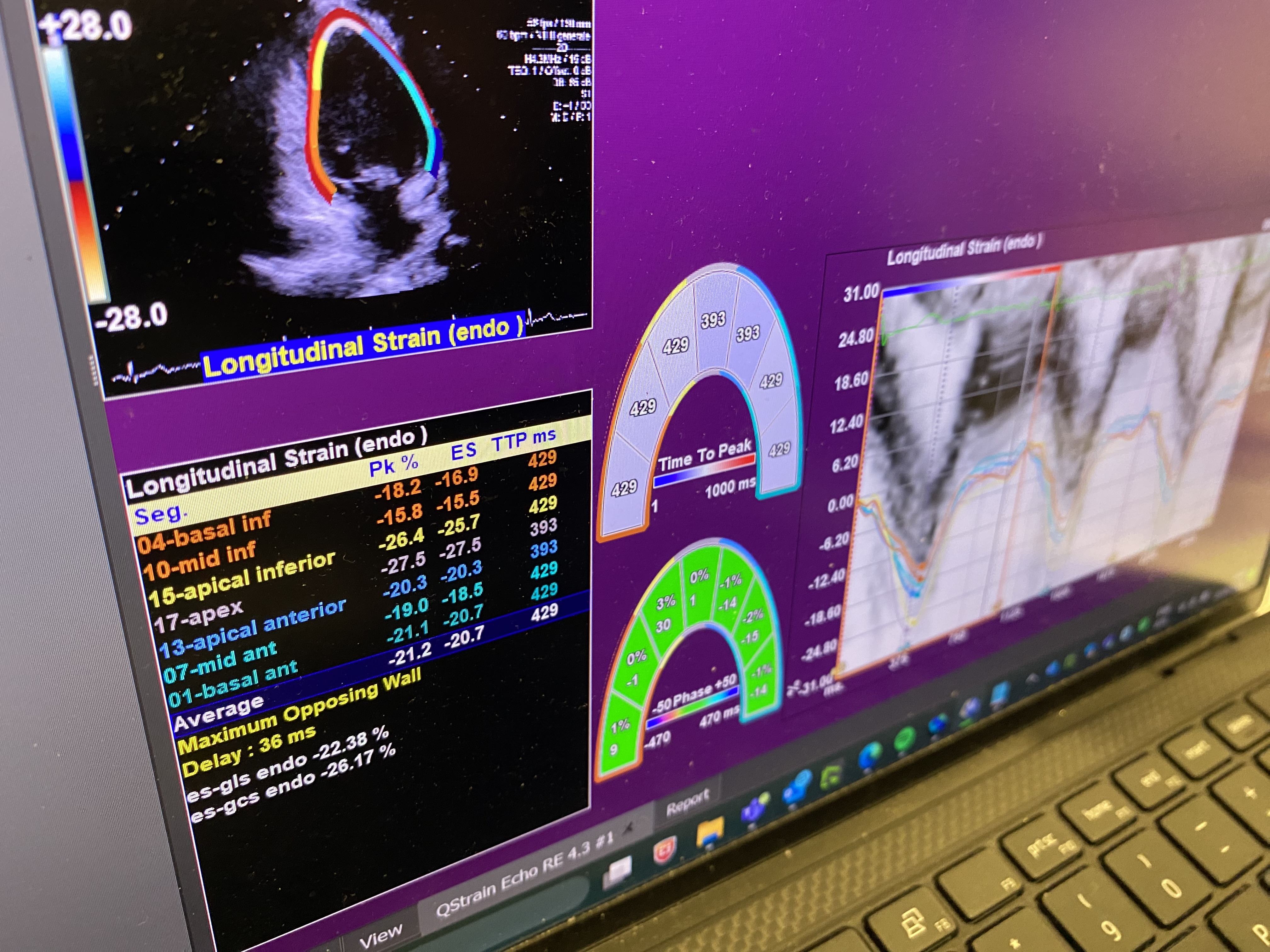
The Medis QStrain Echo software for strain ultrasound.
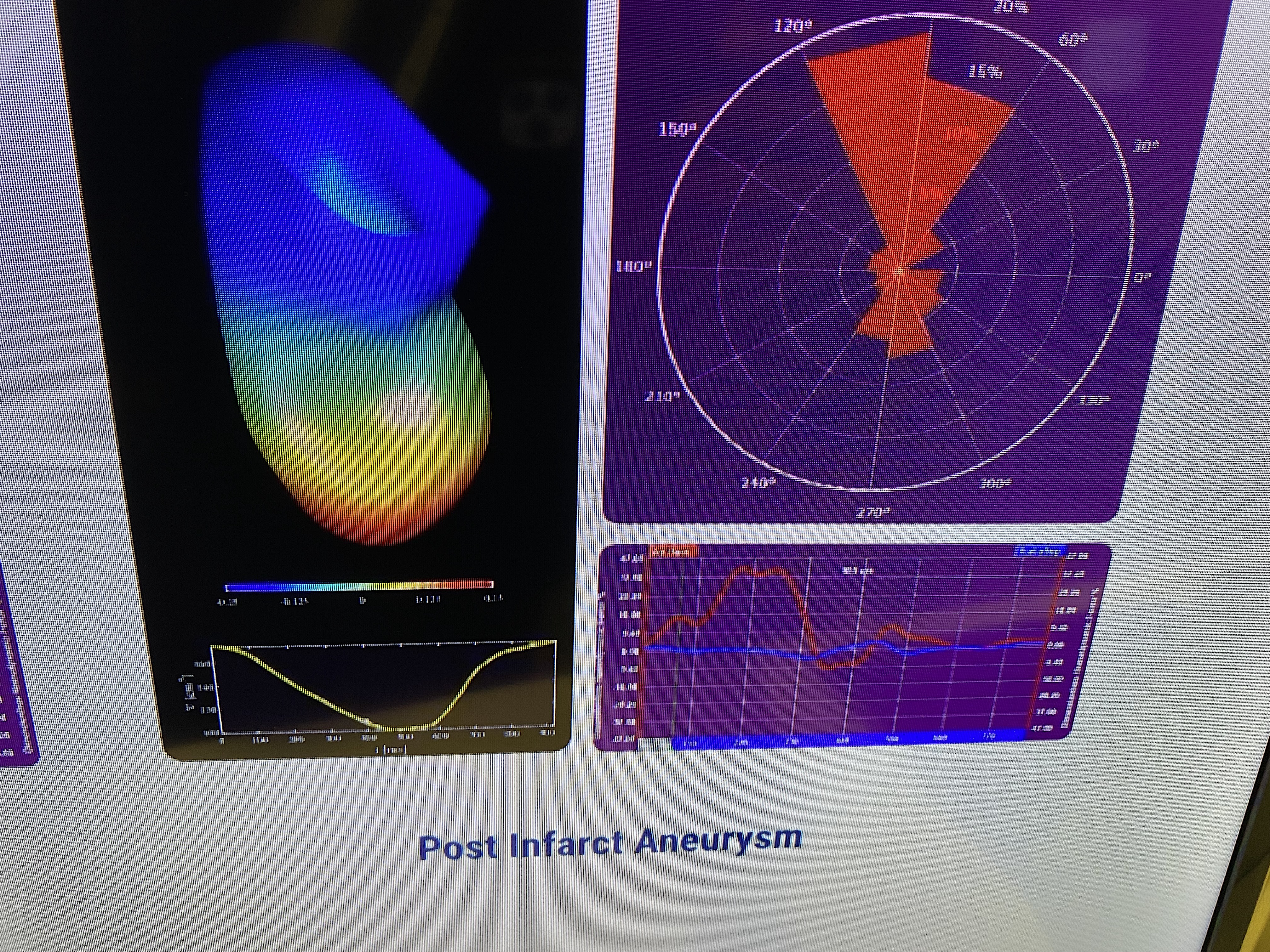
A color-coded map showing strain patterns in a 3D format to help identify heart diseases based on these maps using the Medis QStrain Echo software.

ASE attendee playing with puppies at Bark Park on expo floor ASE23. These dog parks on the show floor run by local animal shelters and rescues have become very popular at recent medical meetings, including AHA, HRS, ACC and HIMSS.
US2.AI automated AI assessment for all the measurements needed for a full echo exam in the echo lab or on POCUS ultrasound systems demonstrated on a live patient and available immediately after scanning in a demo at ASE. This AI technology is designed to standardize cardiac ultrasound measurements to enhance accuracy and reproducibility. The technology also can save a great deal of time during and post-processing these exams.
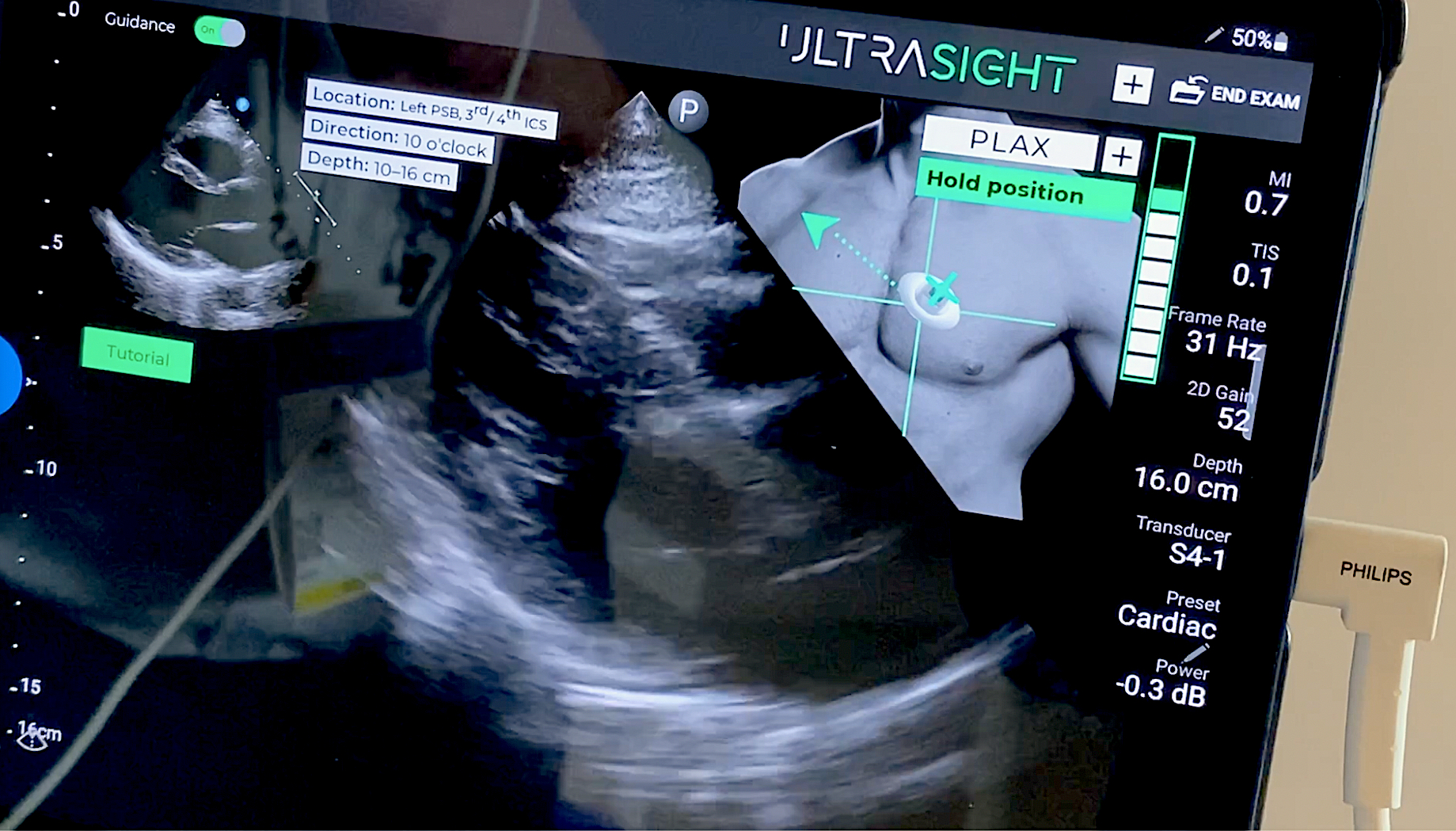
Example of a live POCUS cardiac scan with AI guidance technology to help the user get diagnostic quality echos using the Ultrasight AI system. The AI uses a thumbnail image of the chest and transducer with a target area marked to show where and how to move the transducer. A bar graph on the right side of the thumbnail image shows the quality of the image being acquired and will go up as the user gets into a more ideal imaging window with the correct anatomy in frame.
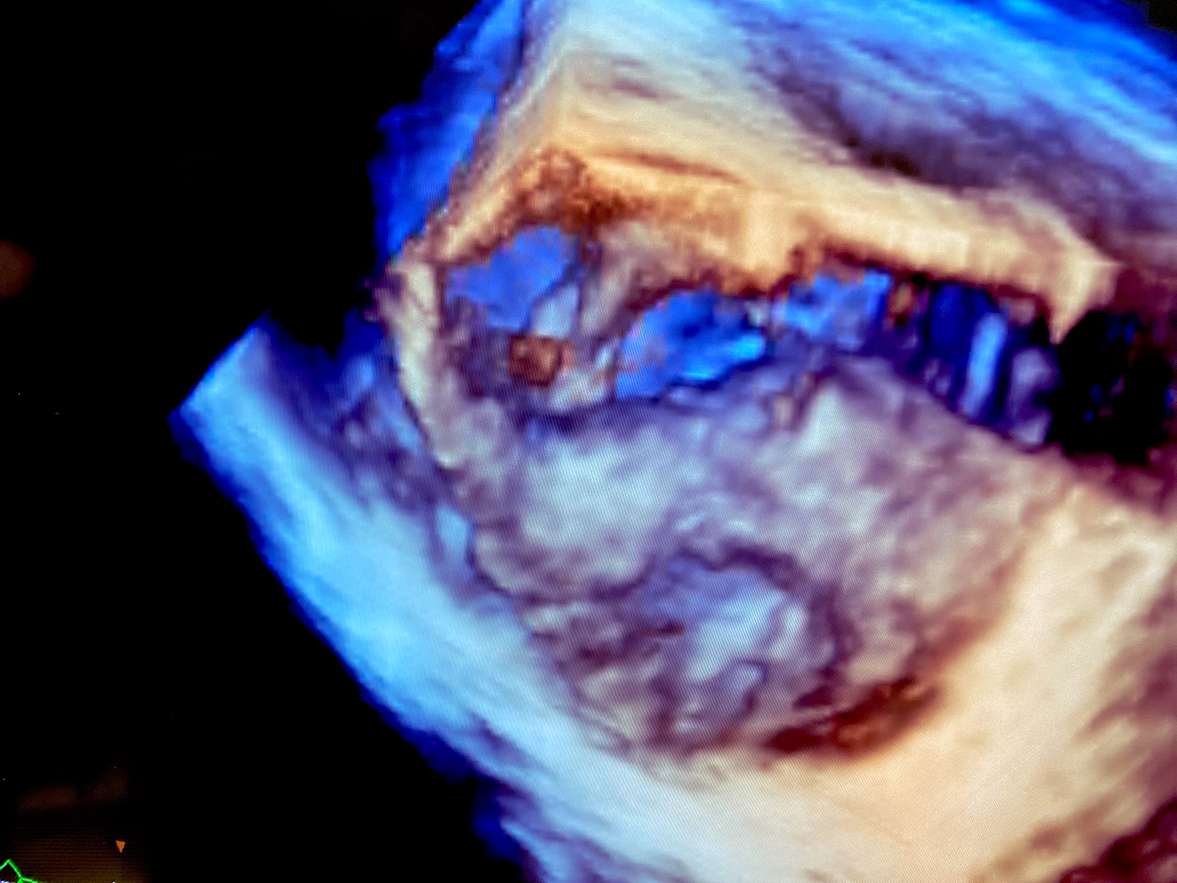
An example of 3D ICE showing a vegetation clot formed on a valve using the Philips ICE system in a demo at ASE.
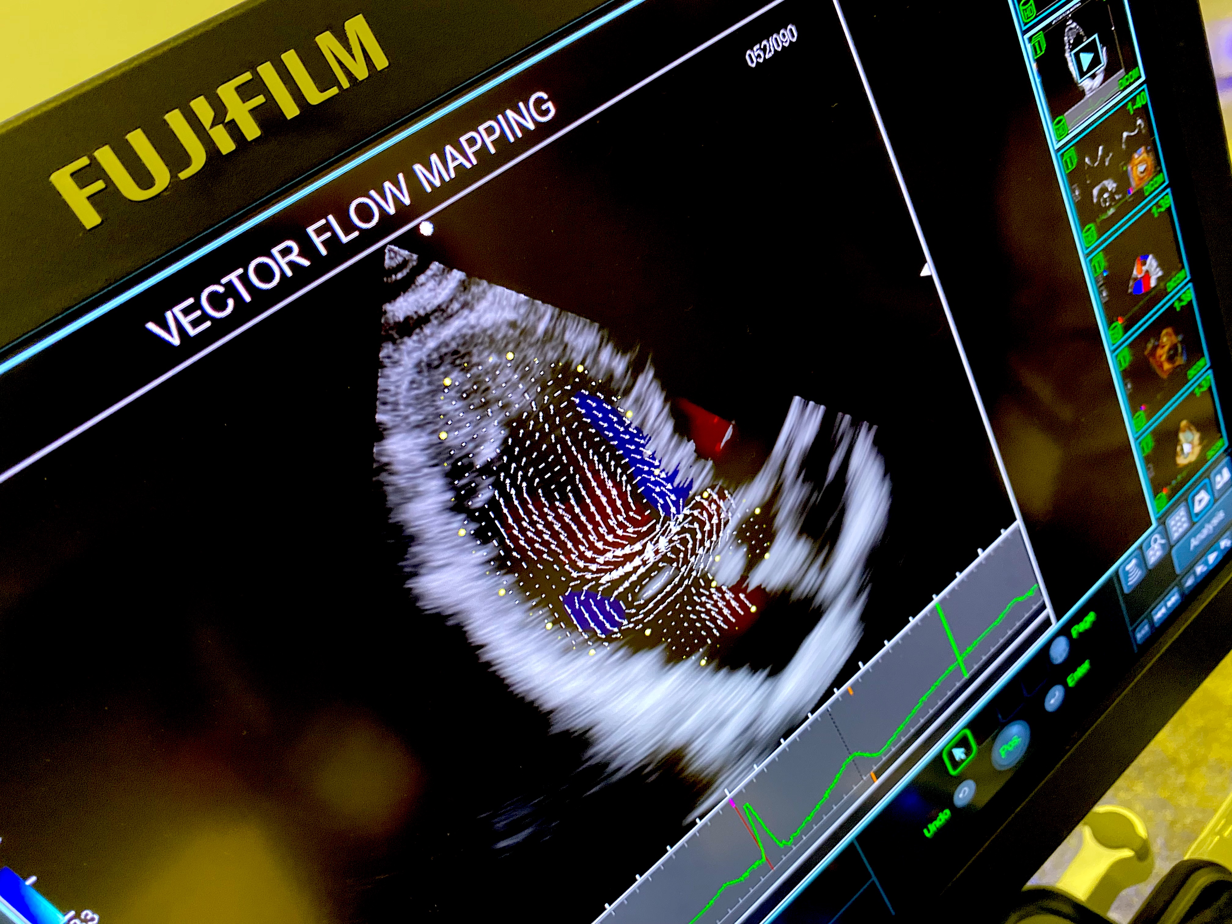
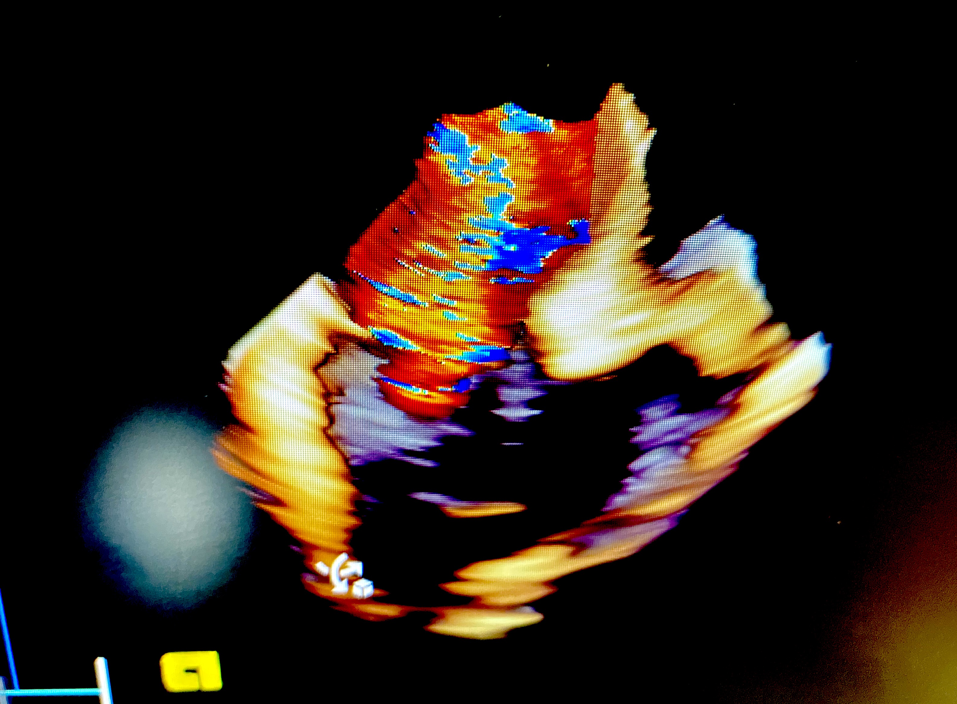
Example of vector flow imaging from cardiac ultrasound, which can track the flow of individual blood cells in the heart and reveal flow patterns. This image is from the Fujifilm Arietta 750 Cardiovascular ultrasound system, but a couple vendors now have similar ultrasound blood tracking technologies. It is believed by many cardiology experts that this type of visualization may be able to detect disease or progression of disease long before a patient becomes symptomatic and one day help better risk stratify patients, but it is still mainly in the research stage.
Example of mitral valve regurgitation seen on 3D ICE using the Siemens technology.
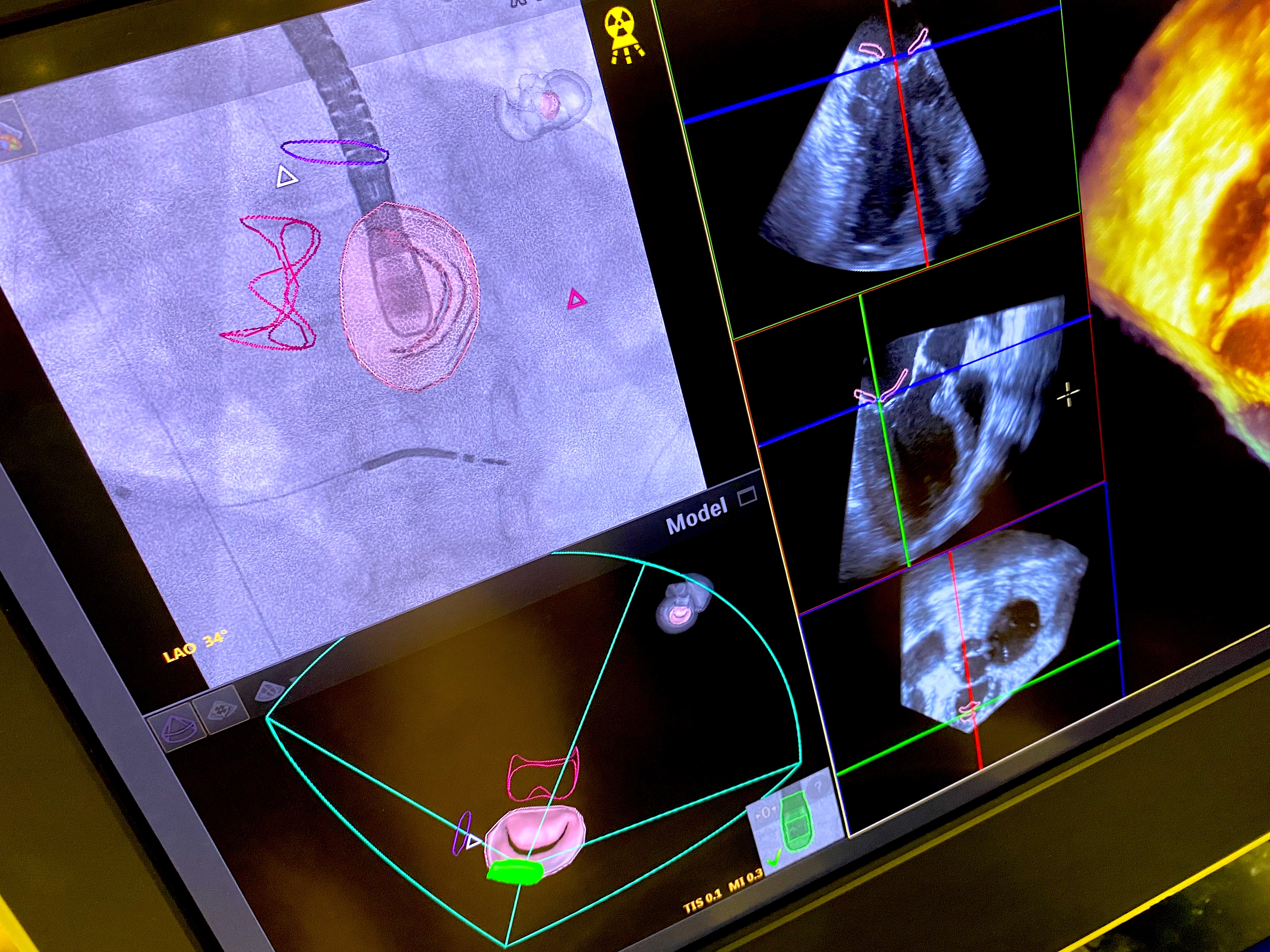
Example of live echo-angiography fusion imaging showing the Mitral valve on TEE using the EchoNavigator technology offered by Philips. This technology helps guide structural heart procedures.
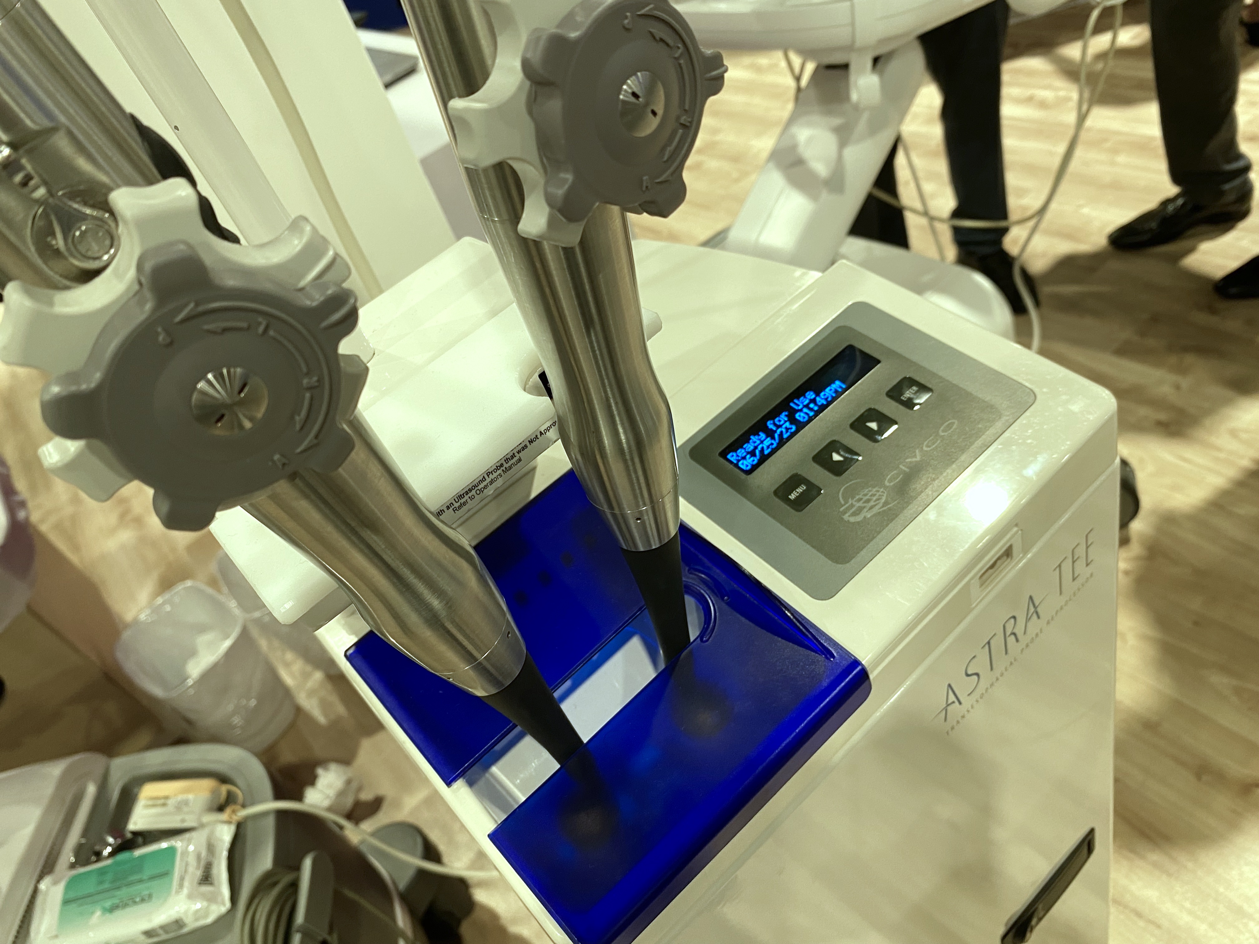
Astra TEE sterilizer in Philips Booth.
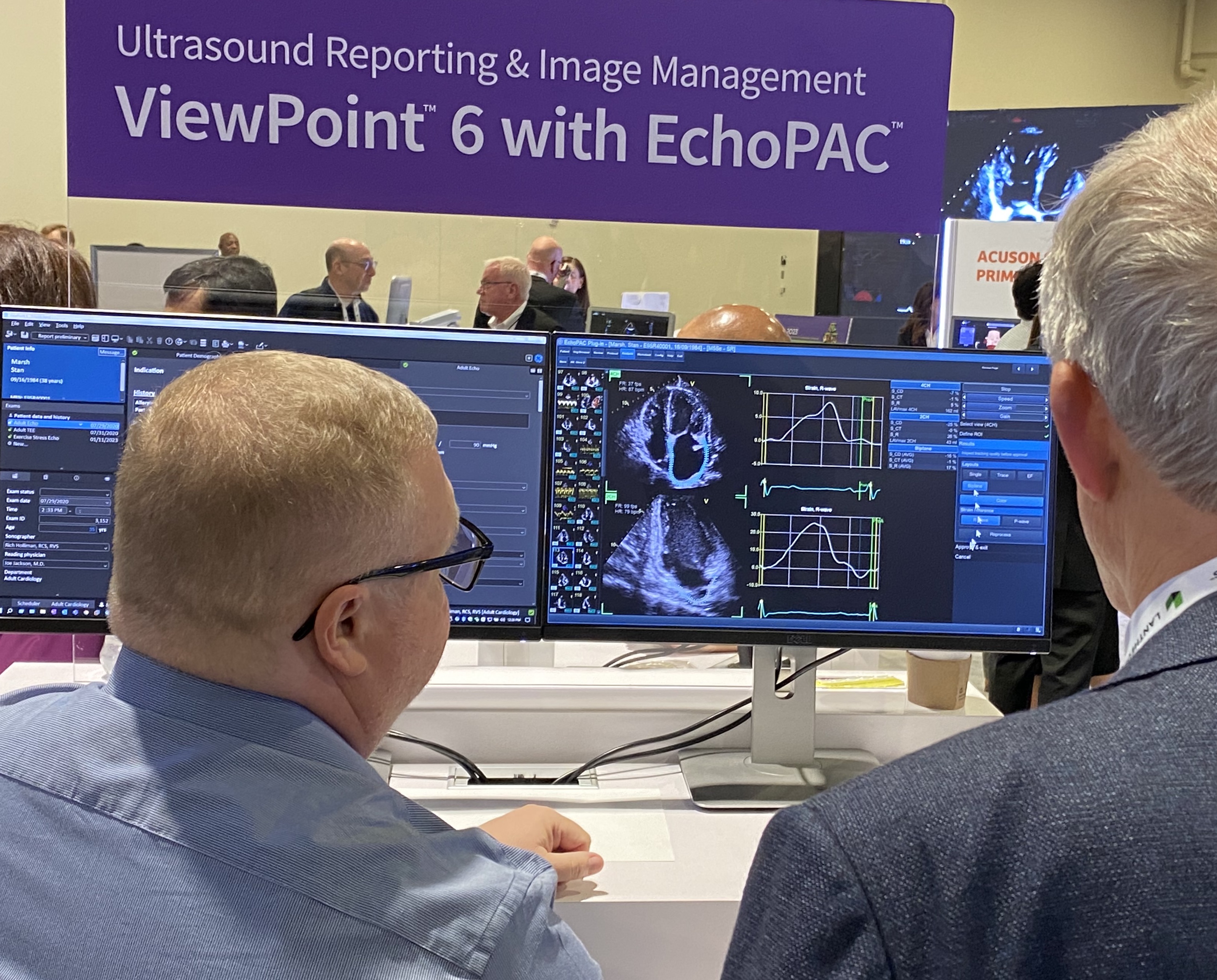
GE Centricity ViewPoint echo reporting CVIS system demo.
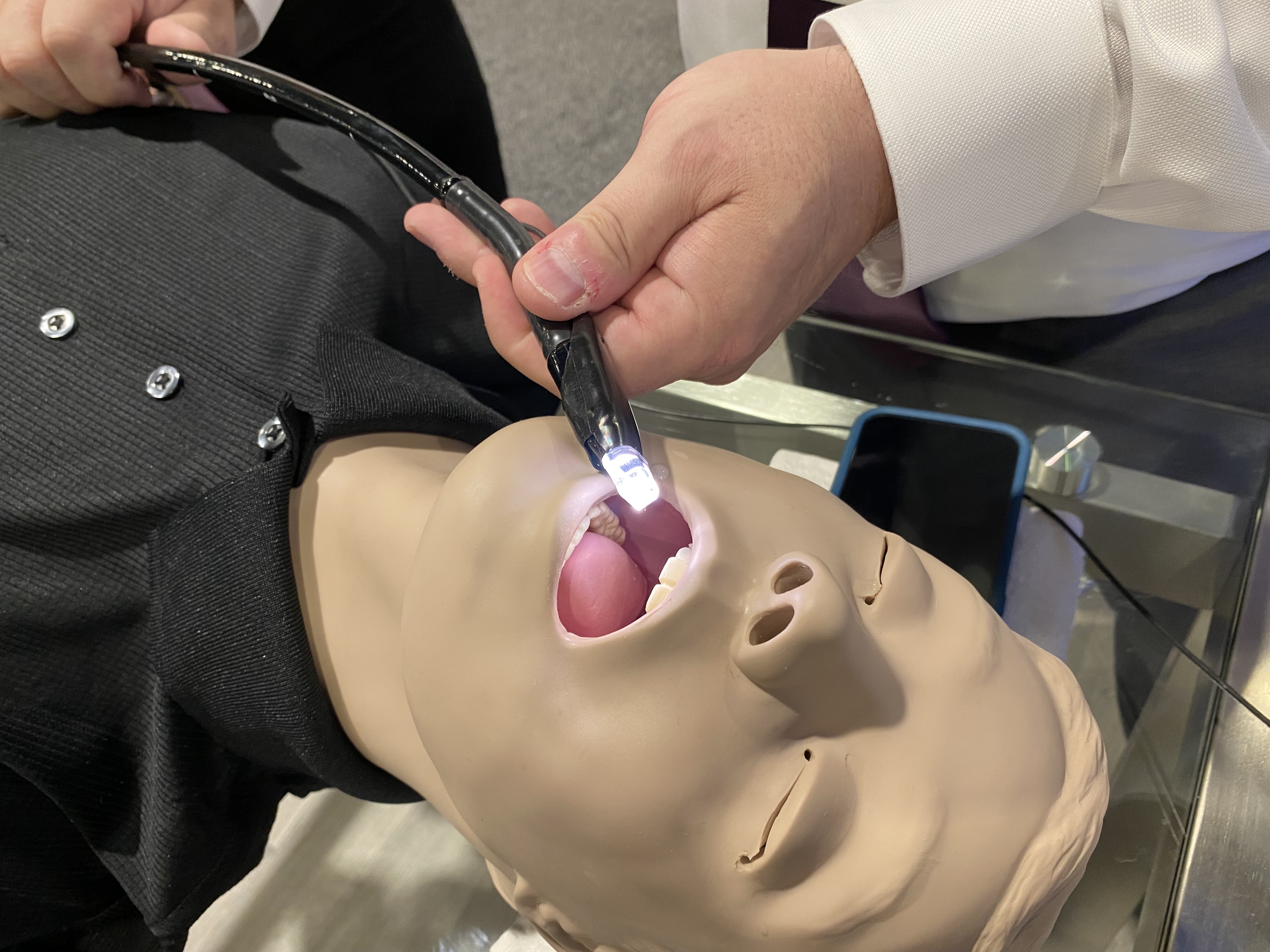
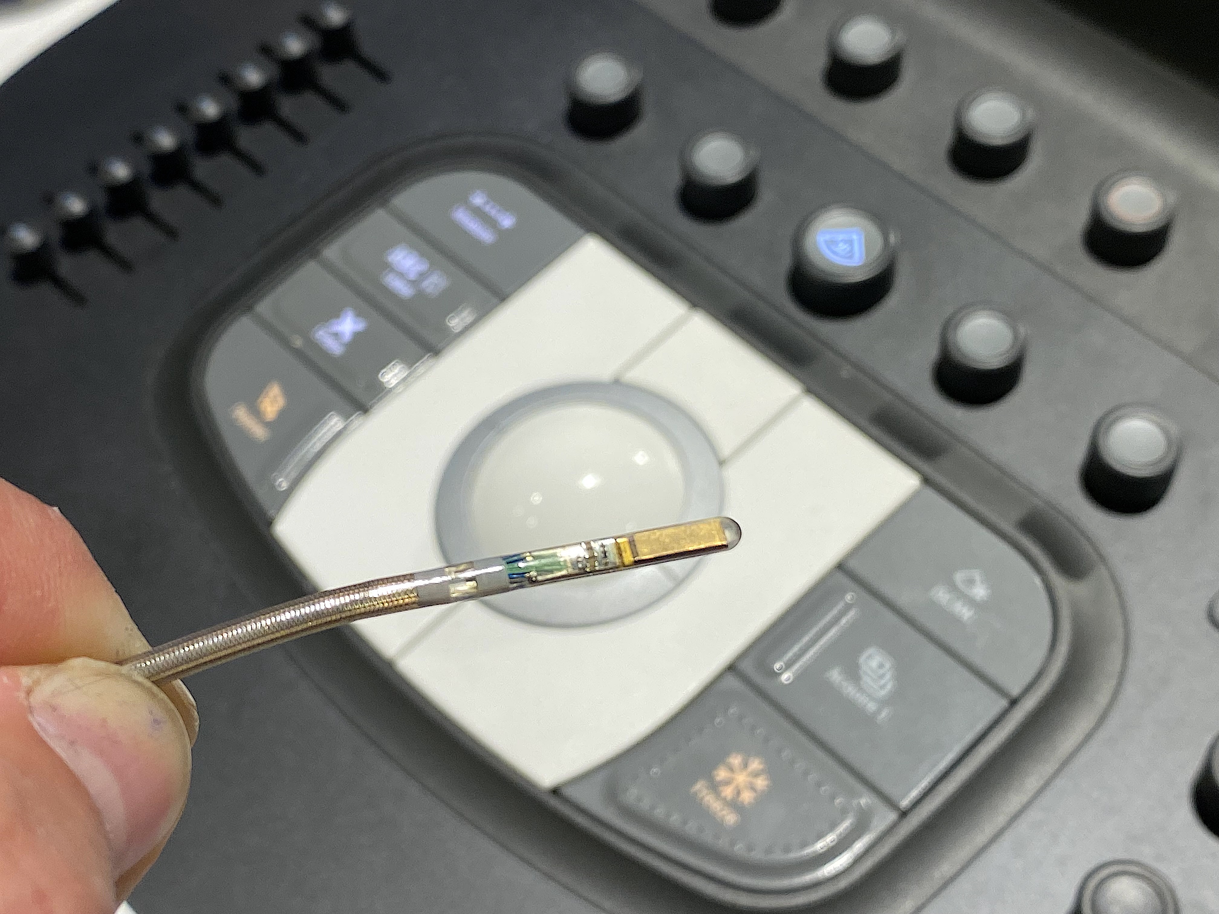
This is the Visura image guided TEE probe tip. It includes a camera and light that clips onto an existing TEE probe so the echocardiographer can can see where the probe is going in the esophagus to avoid injuries that can lead to major complications. It was designed by an interventional echocardiographer because of his concerns about complications.
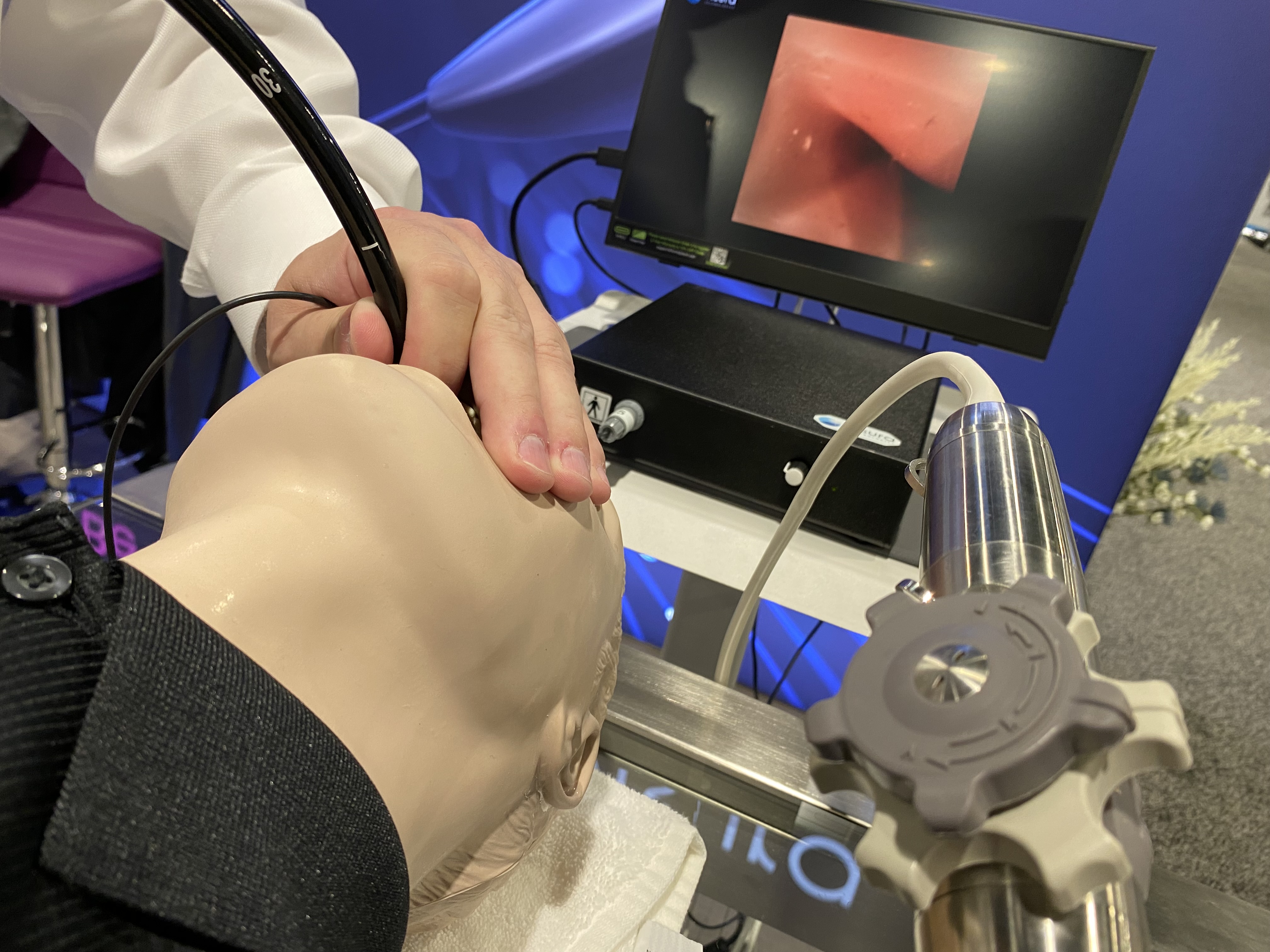
This is the Visura image-guided TEE probe tip. It includes a camera and light that clips onto an existing TEE probe so the echocardiographer can can see where the probe is going in the esophagus to avoid injuries that can lead to major complications. It was designed by an interventional echocardiographer because of his concerns about complications.
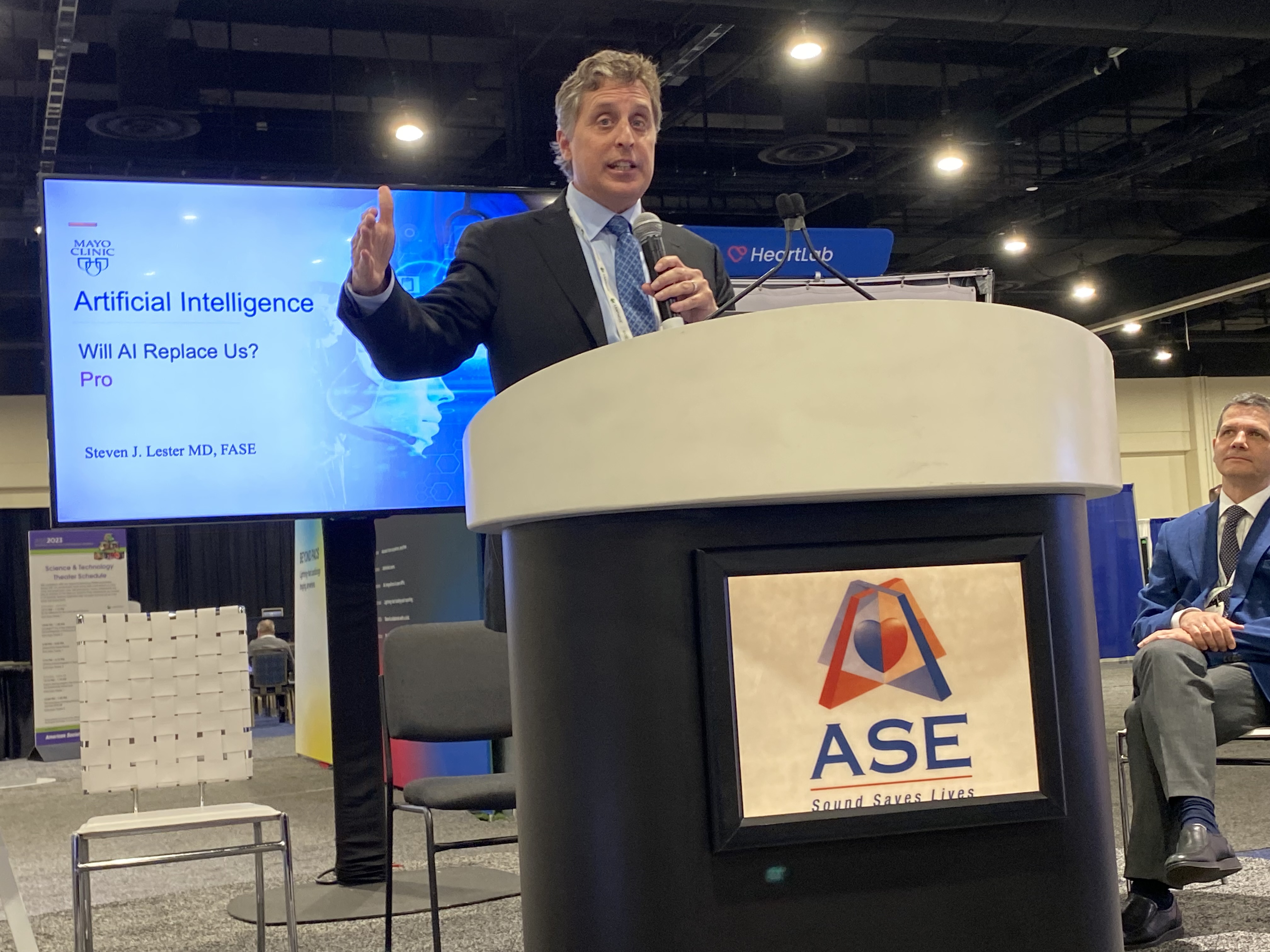
Steven Lester, MD, Mayo Clinic, explains how AI is taking over some roles in the echo lab at an ASE 2023 session on the expo floor. He said automation of measurements, strain and other workflow improvements can save a lot of time and allow clinicians to spend more time focused on the patient rather than using calipers or making lines on a screen.
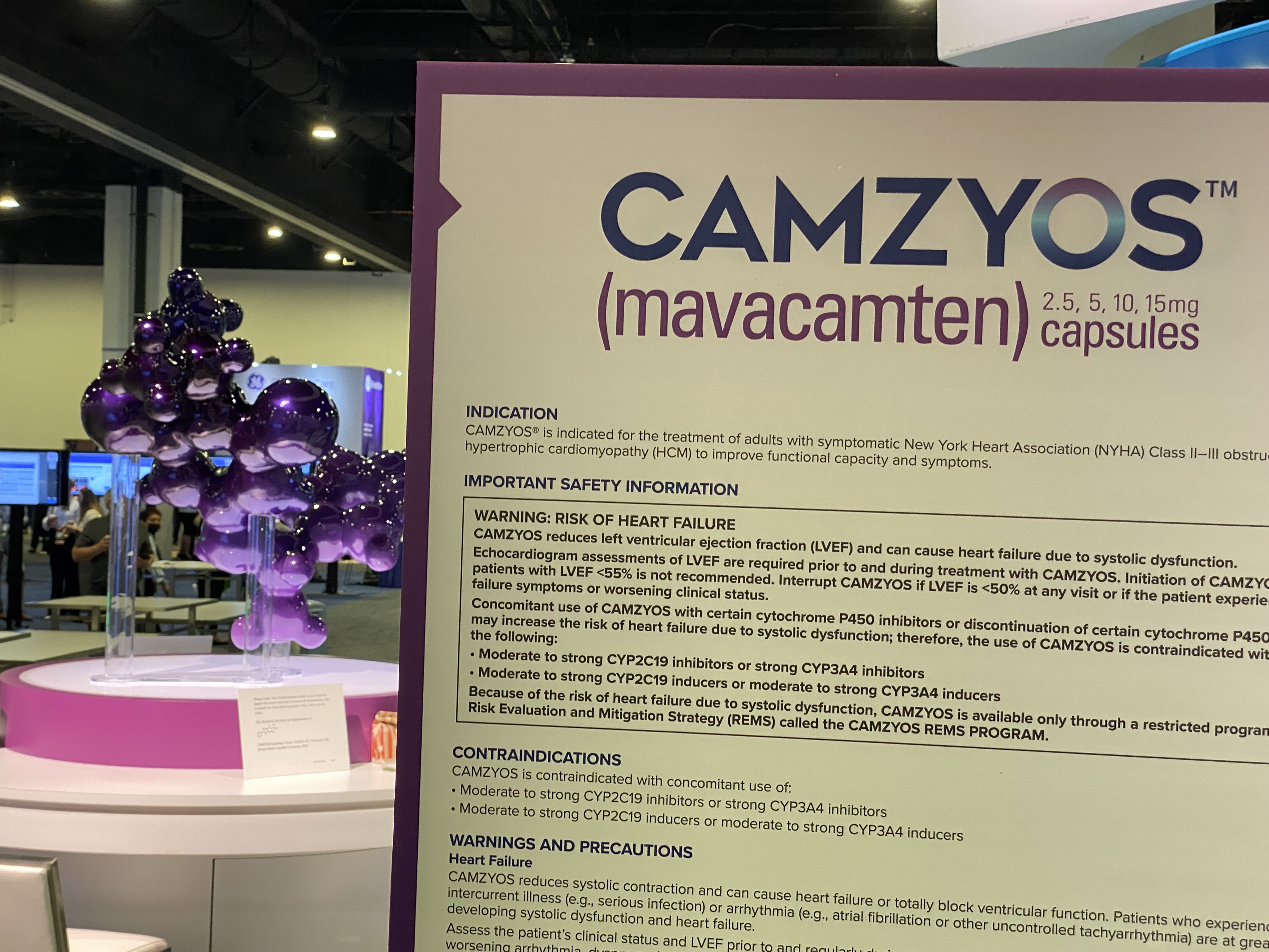
This year at ASE, two pharma vendors had booths for new medications on the market to treat amyloid and mavacanten to treat hypertrophic cardiomyopathy. It is unusual to see drug vendors at ASE, but echo plays a critical role in diagnosing and tracking these diseases. As a result, the drug vendors have become more involved in echocardiography meetings.
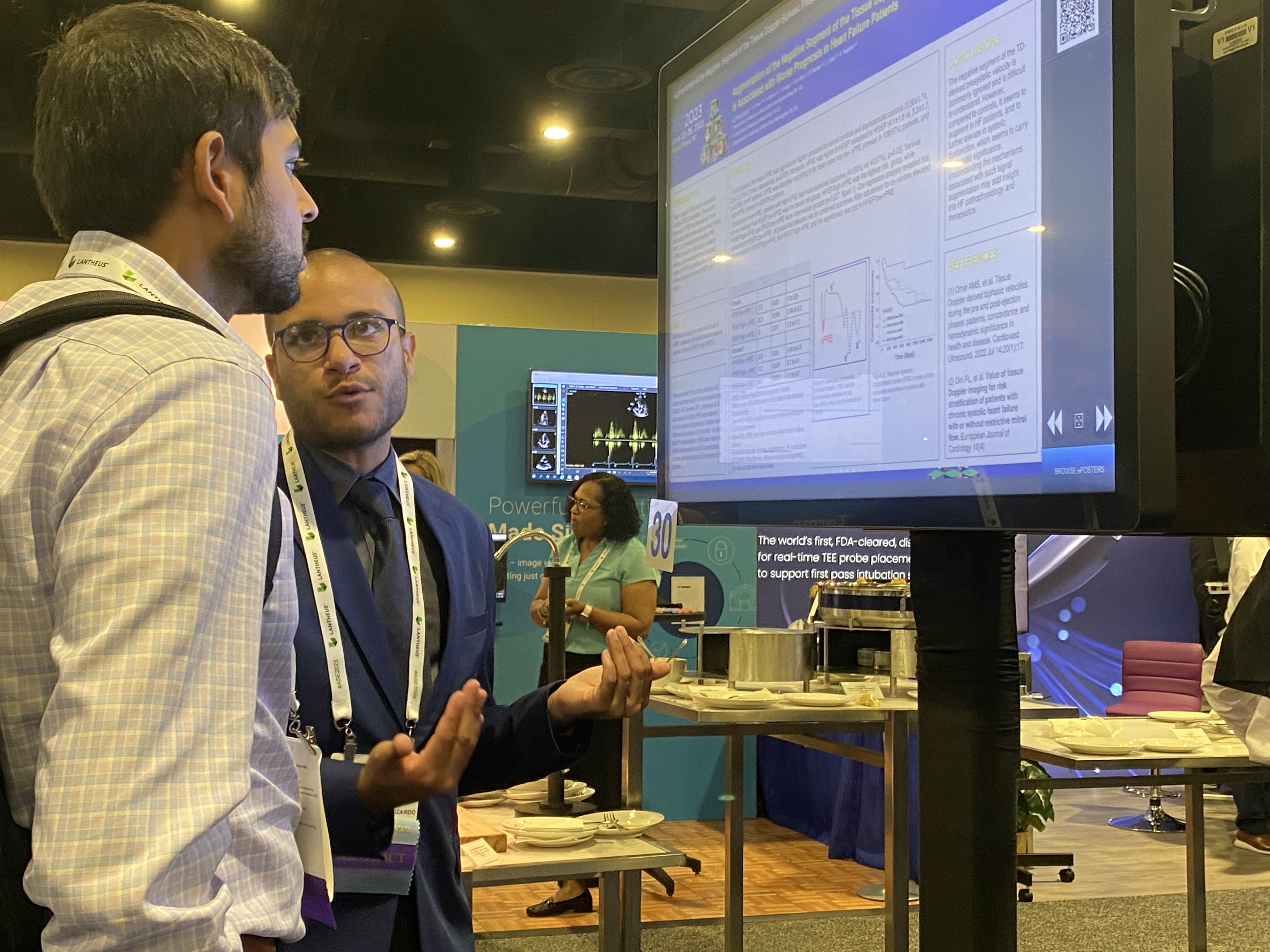
ASE attendee listens as a researcher explains their poster on the expo floor at ASE 2023.
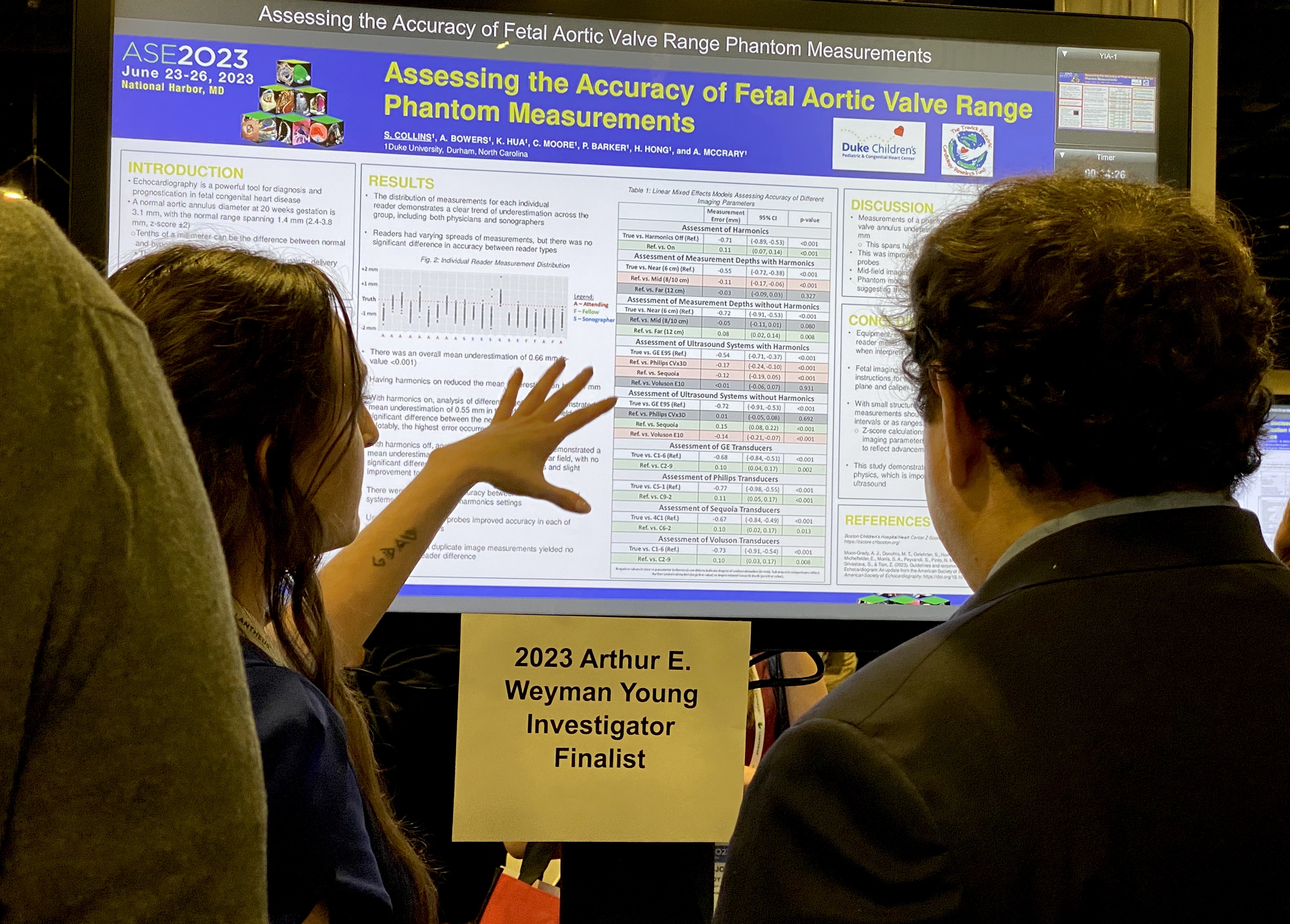
ASE attendee listens as a researcher explains their poster on the expo floor at ASE 2023.
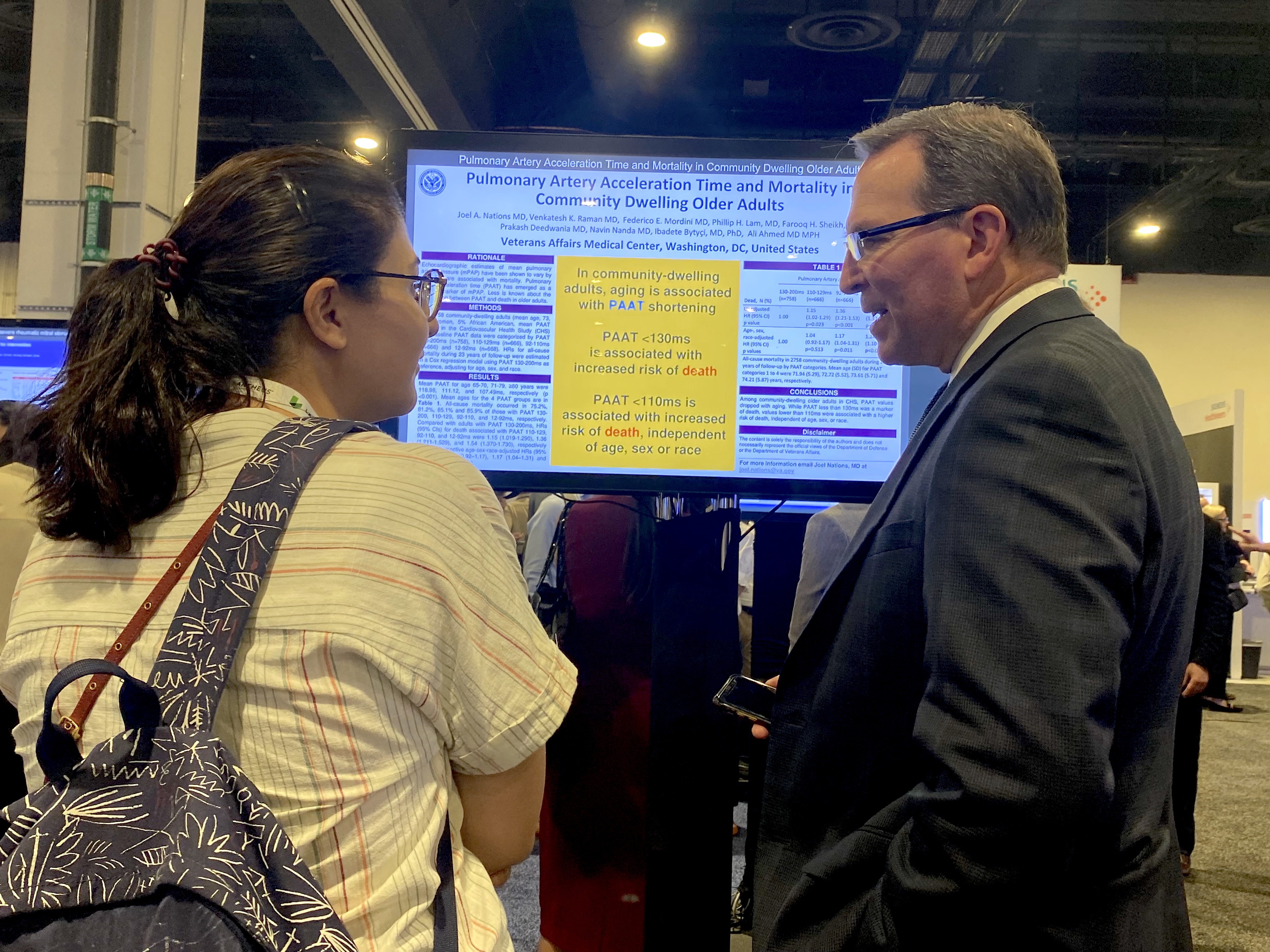
ASE attendee listens as a researcher explains their poster on the expo floor at ASE 2023.
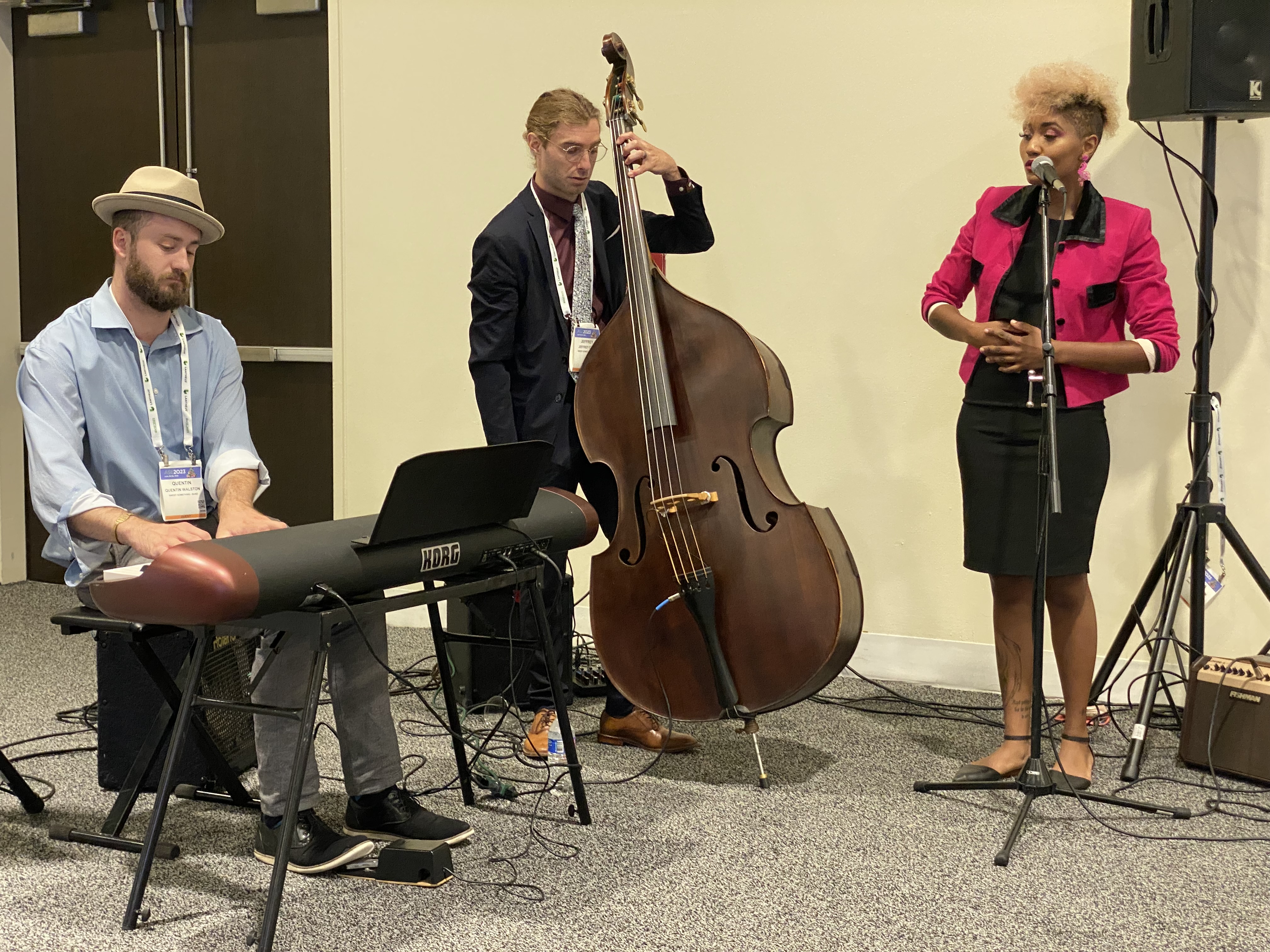
Band playing at during the president's reception on the show floor at ASE 2023.
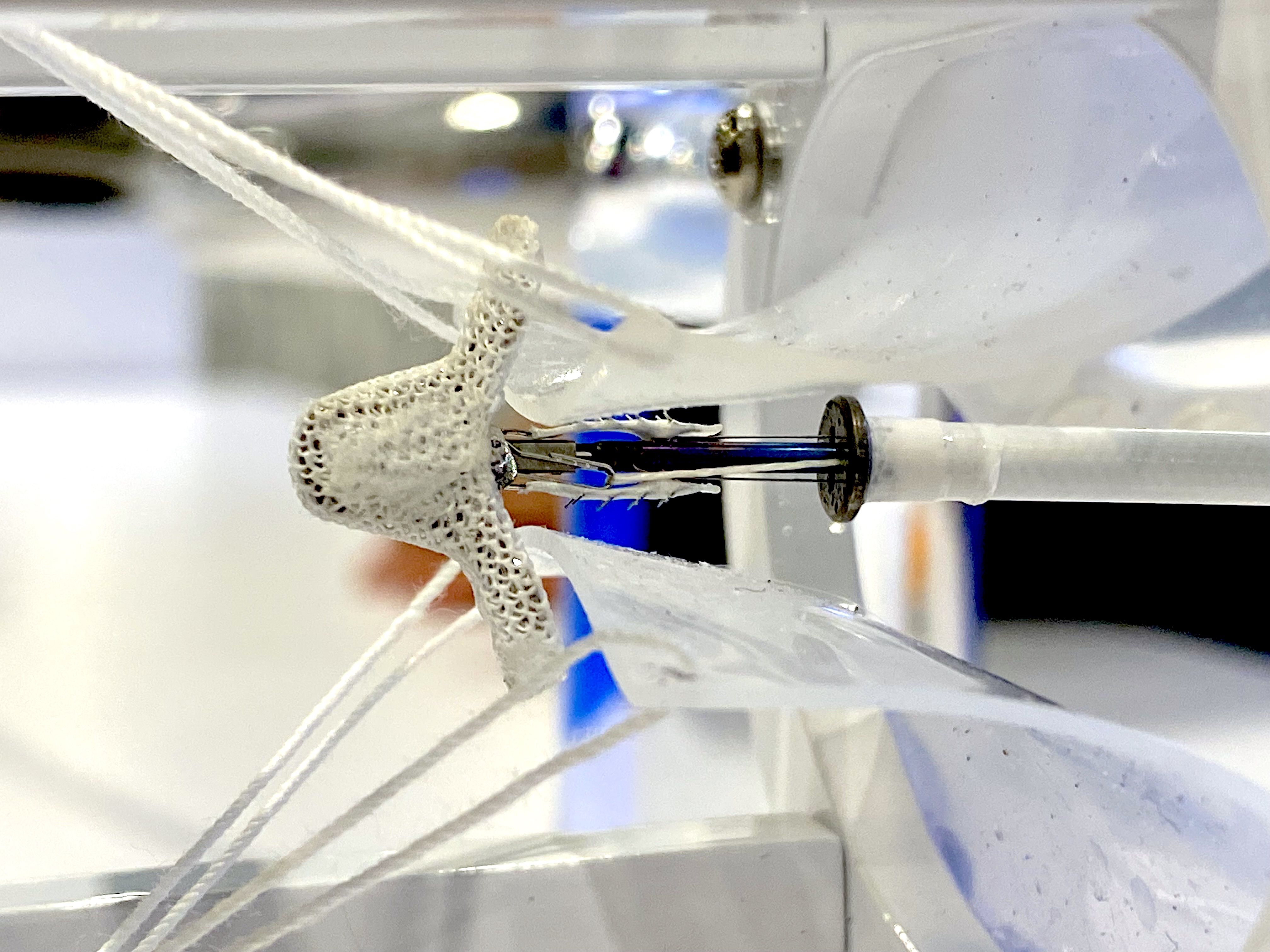
The Abbott MitraClip G4 in a simulated mitral valve for demonstrations at ASE 2023. Abbott felt it was important to set up a booth at ASE because of the critical role echocardiography plays in performing a MitraClip procedure. Without live TEE guidance, these procedures cannot be performed. The display allowed echocardiographers to get an up close view of the clip device and how it operates so they have a better understanding of what they are seeing during procedures.
Example of AI-assisted echo exam guidance on the Echonous Kosmos POCUS system. The thumbnail picture of the chest, target area and transducer show the user how to position and move the transducer to get better quality cardiac images.
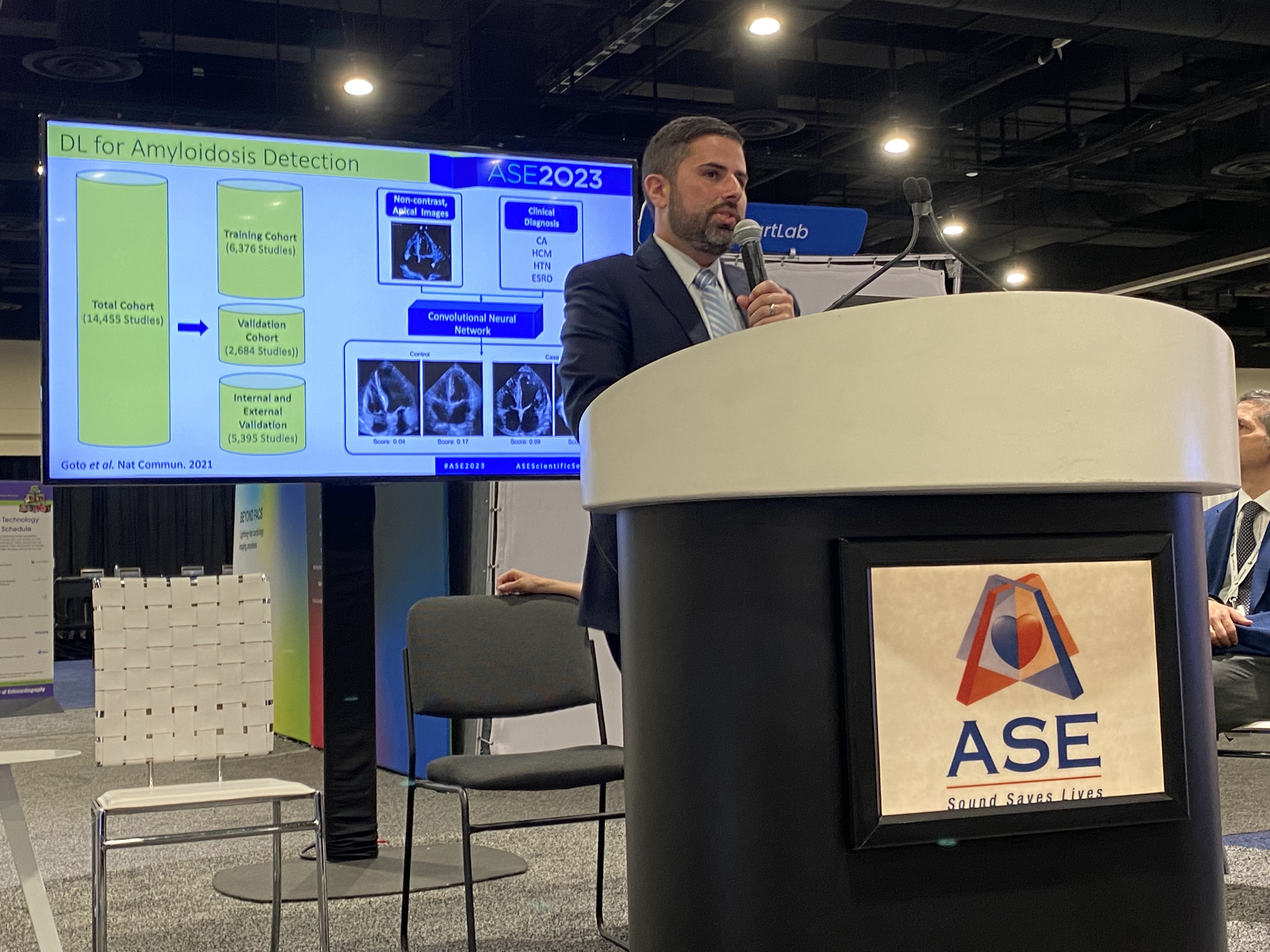
Jeremy Slivnick, MD, University of Chicago, speaks on use of AI for amyloid detection and classification at ASE 2023. At the meeting he was awarded the 2023 Arthur E. Weyman Young Investigator’s Award Competition Winner for his research. The type of amyloid is important to determine for which treatment can be used, but it can be difficult to determine this on echo alone. But, it is expected AI will be used in the coming years to make this differentiation on echo.
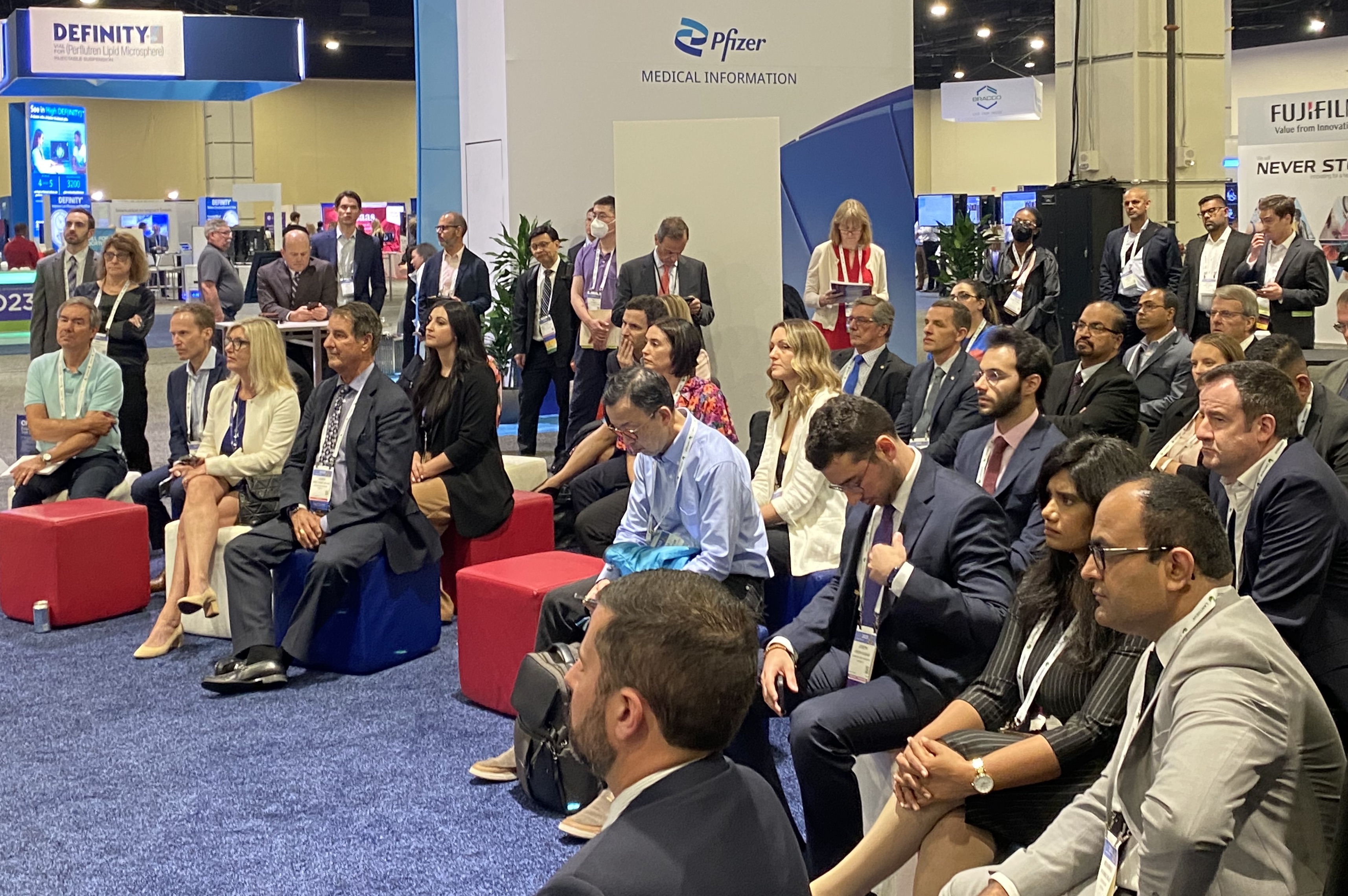
Crowd of attendees listening to one of the AI sessions on ASE23 expo floor.
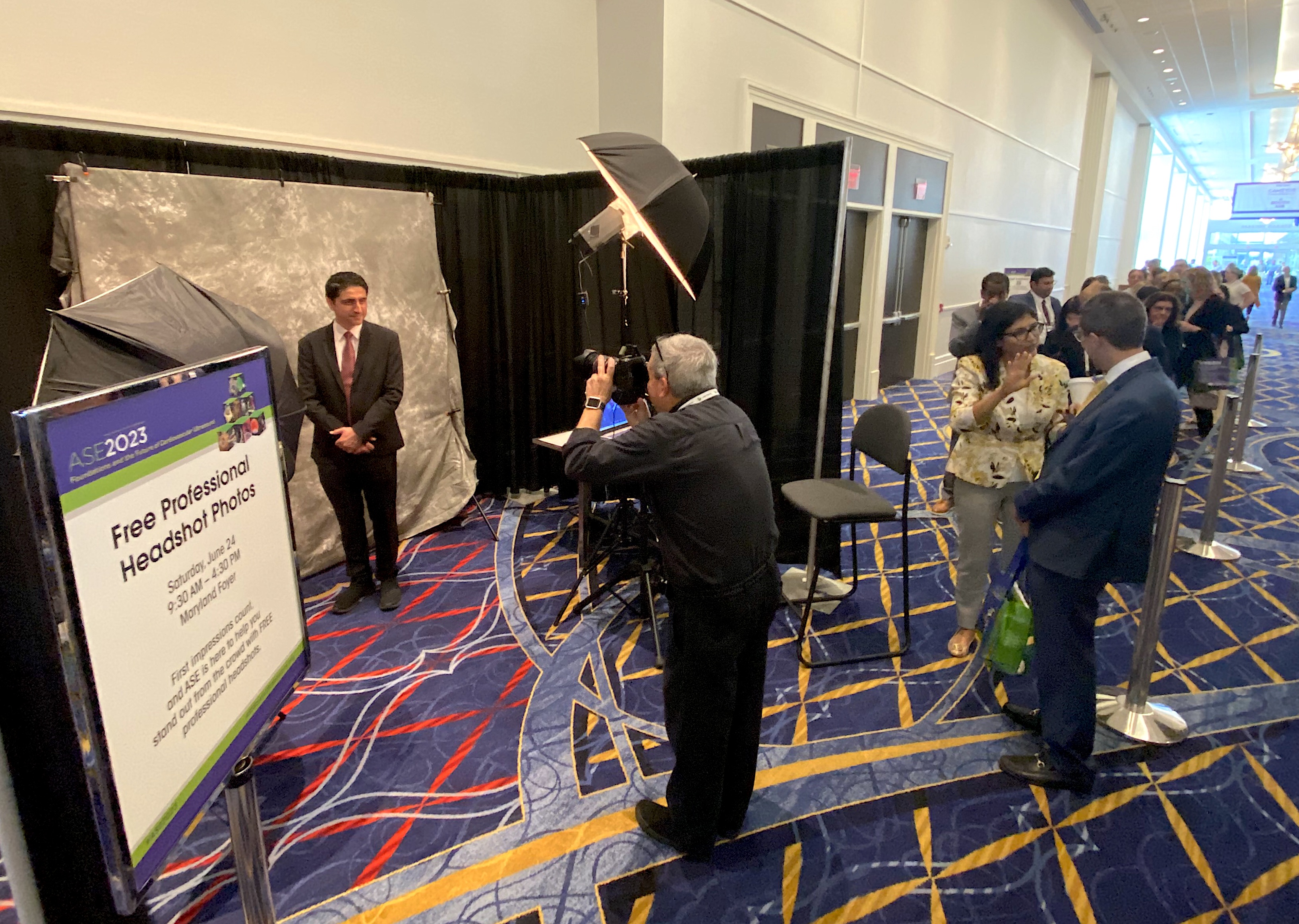
A long line of ASE attendees waiting to have their free professional headshot taken. ASE noted that there is a lot of movement of sonographers going to new jobs during this post-COVID era of the Great Resignation.
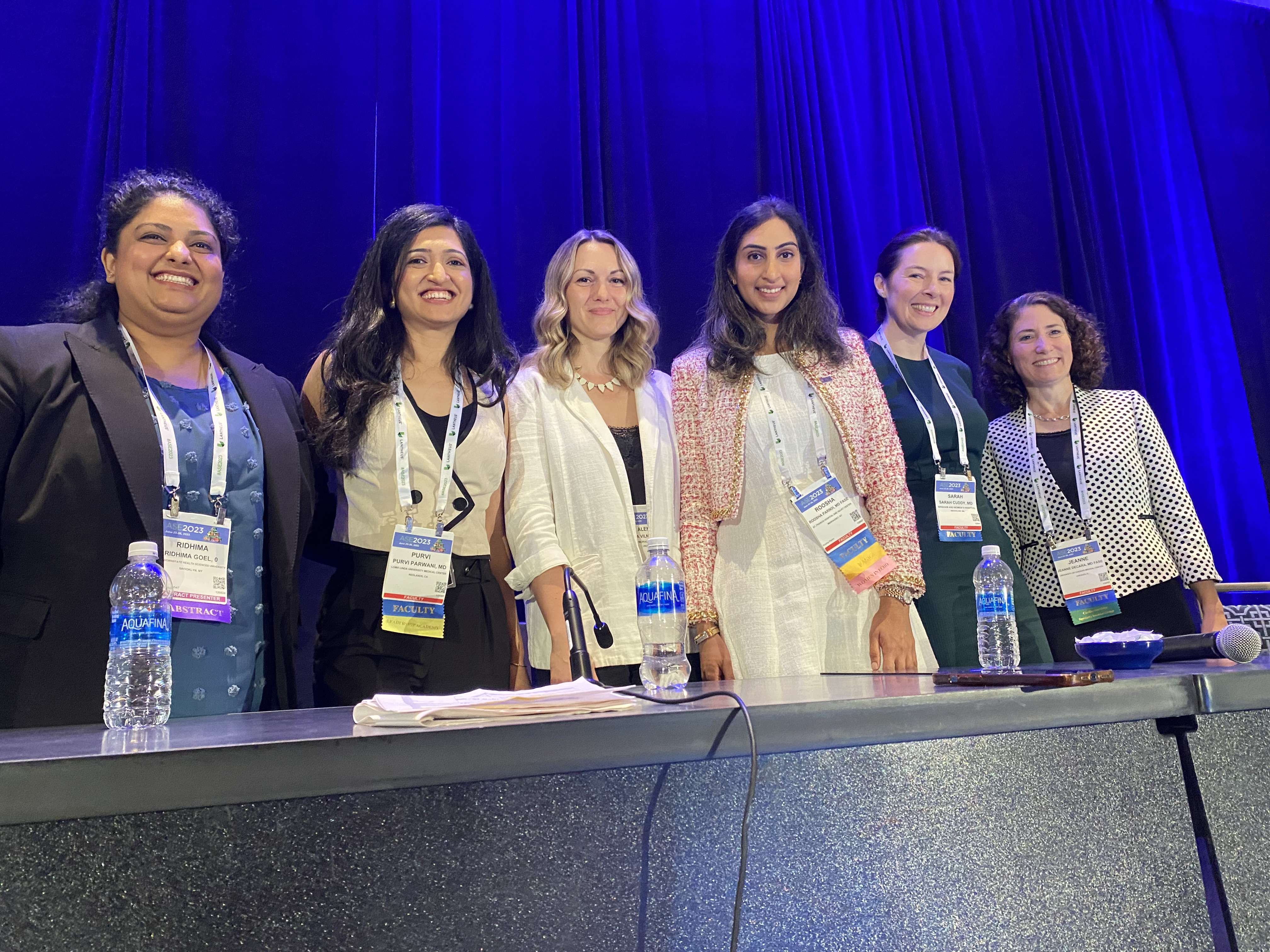
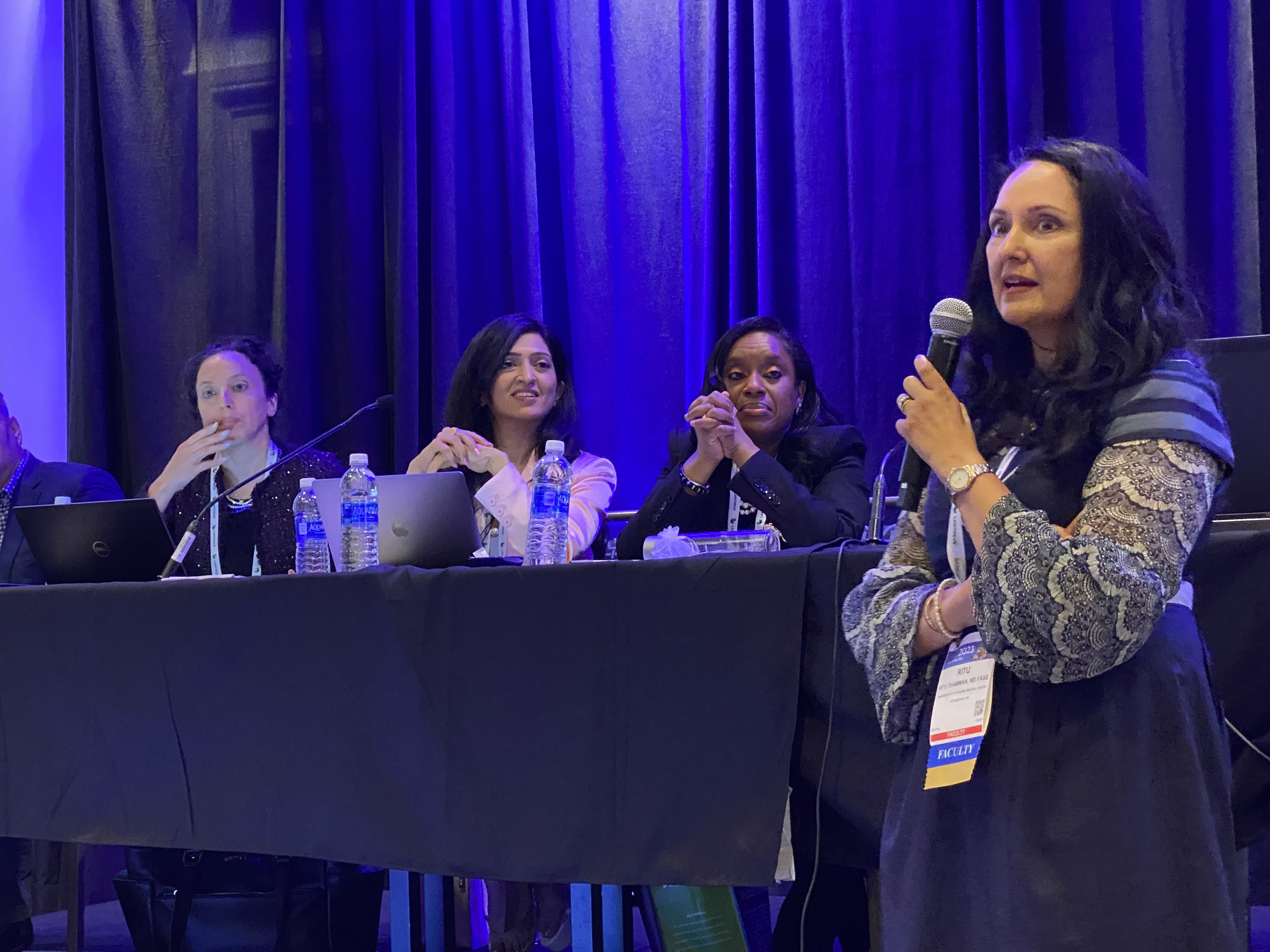
A rare all-women panel at an ASE session focused on amyloid imaging at ASE 2023. From left to right are Ridhima Goel, MBBS, SUNY Downstate Medical Center; Purvi Parwani, MD, Loma Linda; Alena Vilner, sonographer, UMass Memorial Medical Center; Roosha Parikh, MD, St. Francis Hospital; Sarah Cuddy, MD, Brigham and Womens; and Jeanne Decara, MD, University of Chicago.
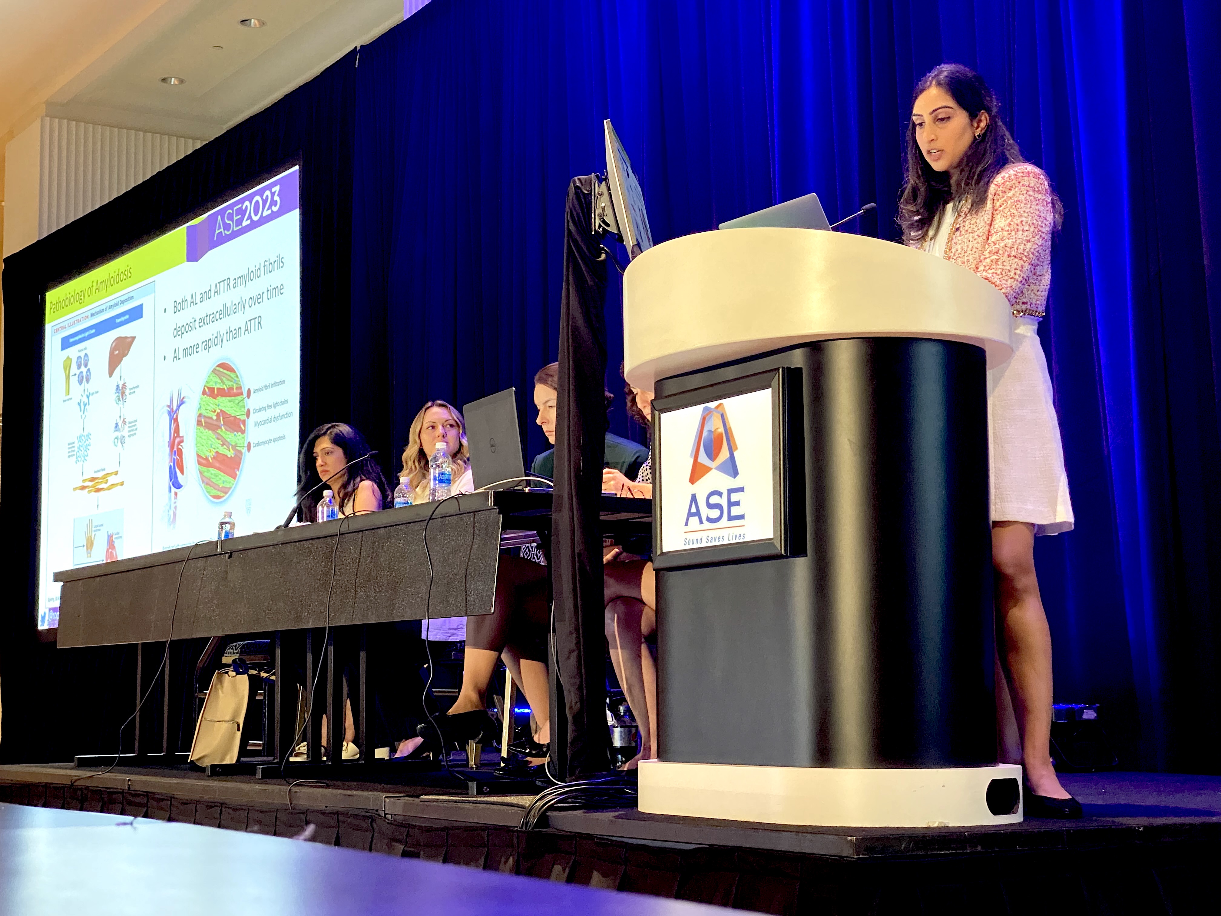
Roosha Parikh MD, with St. Francis Hospital in New York, presenting an overview session on the different types of amyloid during ASE 2023. With a new drug now available to treat amyloid, there has been increasing interest to learn how to image for the disease.
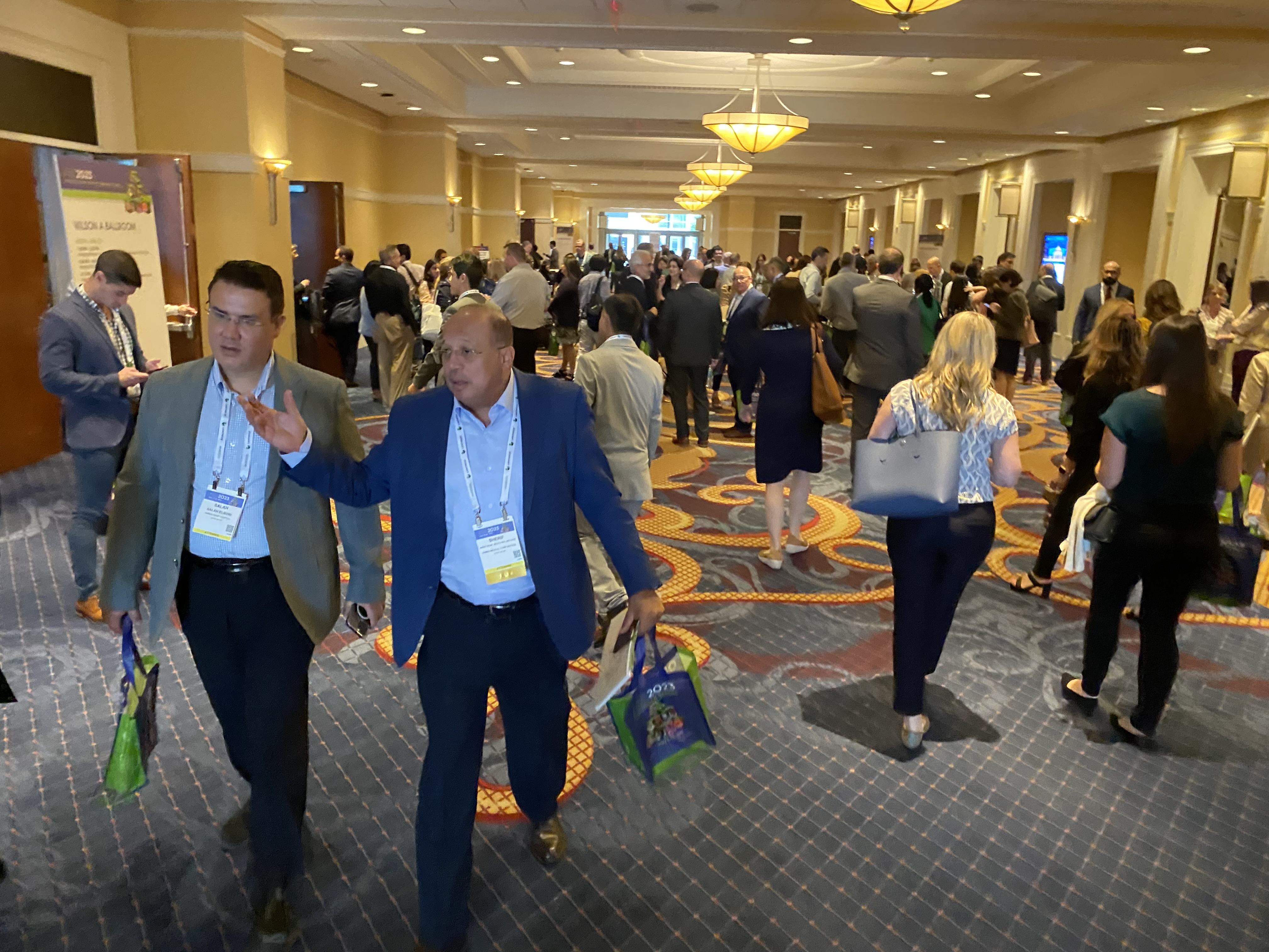
Attendees between sessions at the ASE 2023 meeting.
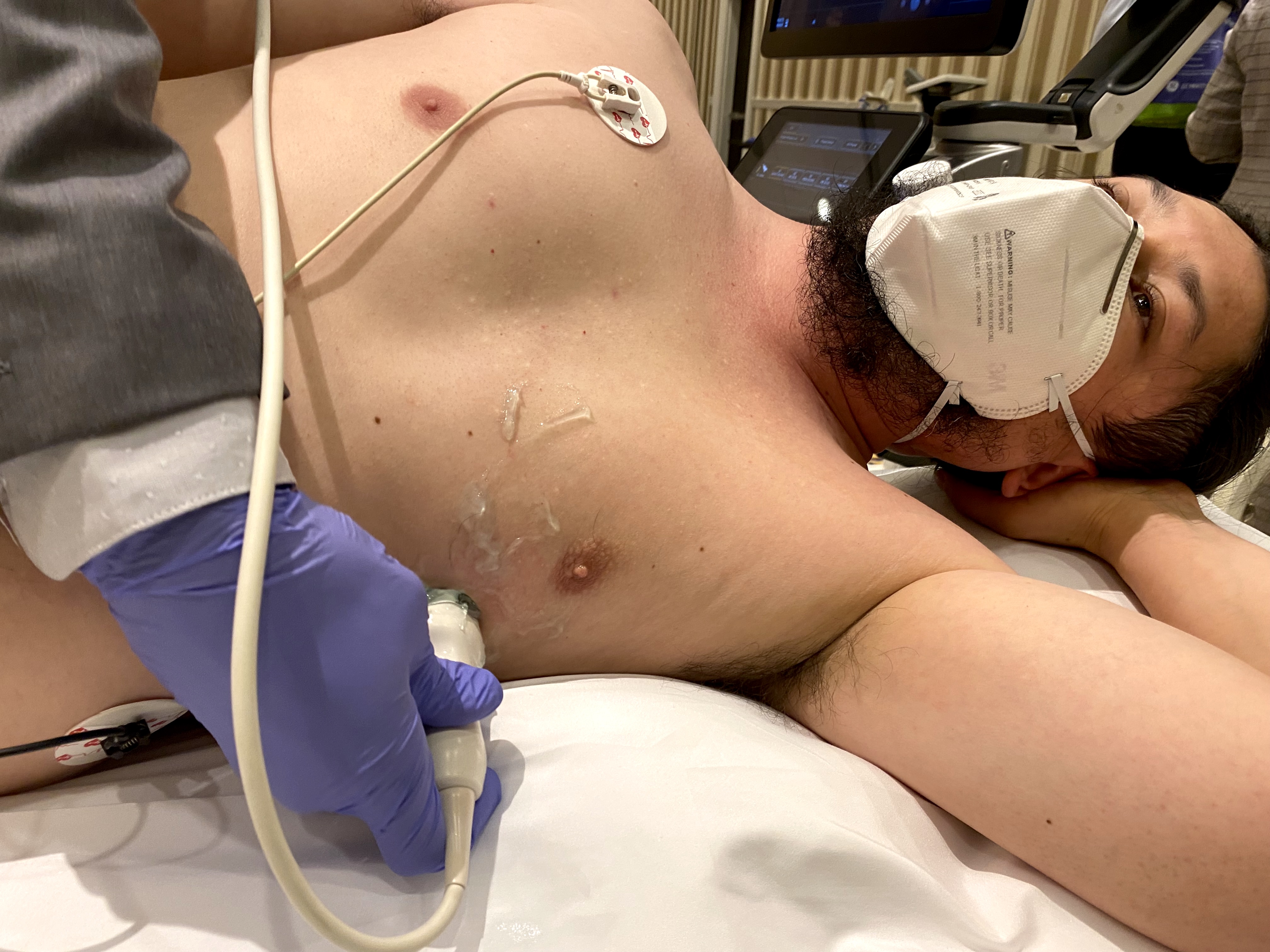
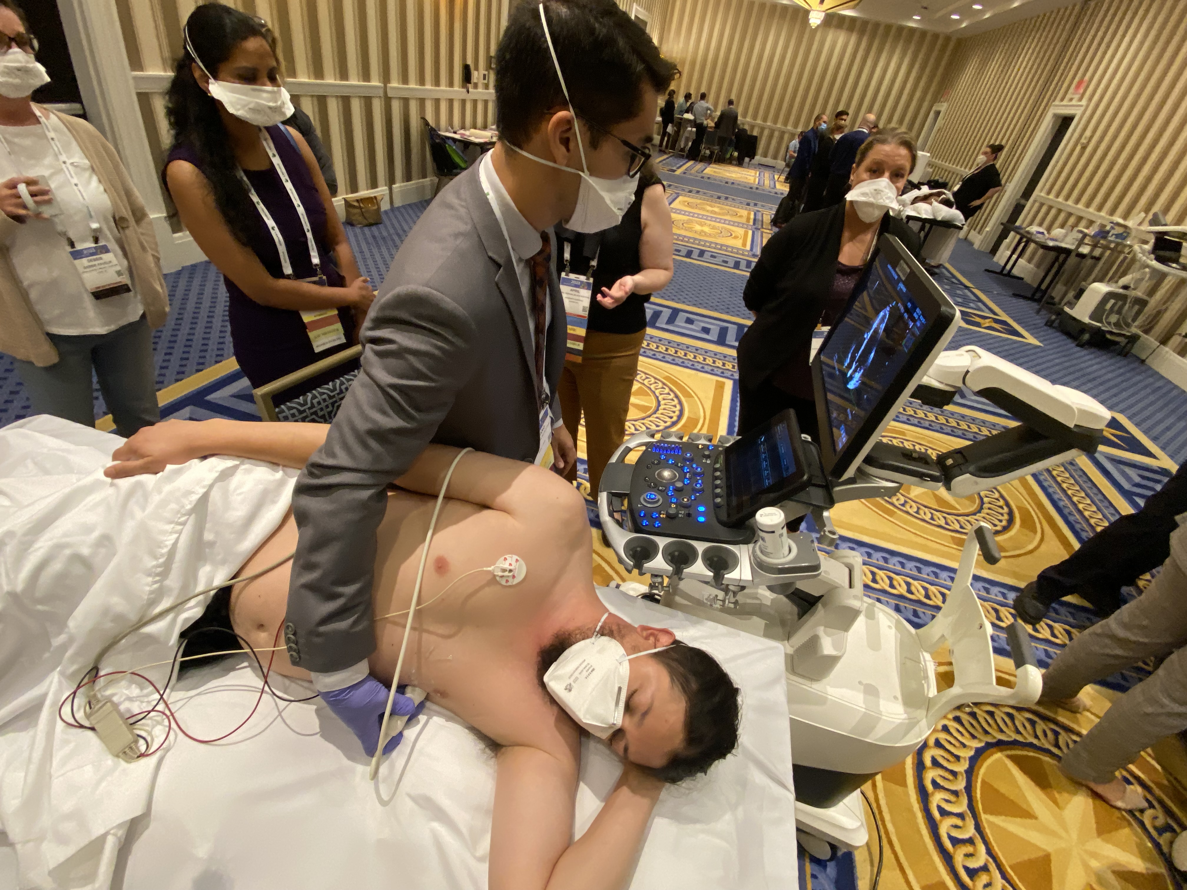
Cardiac ultrasound being performed during a hands-on strain echo training session. Participants were able to try out strain imaging on several vendor's ultrasound platforms and rotated between these vendor stations.
Sonographer Alicia Armour, Heart Center Administrator with Duke Triangle Heart Associates, presenting a patient case in an ASE session on strain imaging. She also presented sessions on HCM and scanning in a hyperbaric chamber. Sonographers make up 45% of the ASE membership and the society includes large numbers of them as presenters in sessions at the annual meeting.

Dave Fornell has covered healthcare for more than 17 years, with a focus in cardiology and radiology. Fornell is a 5-time winner of a Jesse H. Neal Award, the most prestigious editorial honors in the field of specialized journalism. The wins included best technical content, best use of social media and best COVID-19 coverage. Fornell was also a three-time Neal finalist for best range of work by a single author. He produces more than 100 editorial videos each year, most of them interviews with key opinion leaders in medicine. He also writes technical articles, covers key trends, conducts video hospital site visits, and is very involved with social media. E-mail: [email protected]