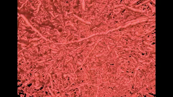
New-look imaging technique could change the way we see blood vessels
Researchers that developed a new imaging technique designed to create full 3D visualizations of blood vessels, shared their progress in Nature Methods.[1]
“Usually, if you want to gather data on blood vessels in a given tissue and combine it with all of its surrounding context like the structure and the types of cells growing there, you have to re-label the tissue several times, acquire multiple images and piece together the complementary information,” corresponding author Arvind Pathak, PhD, professor of radiology, biomedical and electrical engineering at the Johns Hopkins University School of Medicine, said in a prepared statement. “This can be an expensive and time-consuming process that risks destroying the tissue’s architecture, precluding our ability to use the combined information in novel ways.”
The team’s new approach, which they’ve called VascuViz, is designed to make a patient’s blood vessels visible with a variety of different imaging modalities. Instead of only working with only MRI scans or CT scans, for instance, VascuViz helps the blood vessels remain visible at all times.
The team’s work is centered on the combination of a CT contrast agent, BriteVu, and an MRI contrast agent, Galbumin-Rhodamine. This compound makes even tiny details of a patient’s blood vessels visible on multiple modalities. These enhanced images can, in theory, help clinicians learn more about the presence of different blood flow abnormalities and track other changes going on inside the patient’s body.
“Now, rather than using an approximation, we can more precisely estimate features like blood flow in actual blood vessels and combine it with complementary information, such as cell density,” added lead author Akanksha Bhargava, PhD, with the department of radiology and radiological science at Johns Hopkins.
The group’s work is still under development; so far, only tests using mouse tissue have been completed.
Related Vascular and Endovascular Content:
DOACs may reduce the risk of dementia among AFib patients by 50%
PCI boosts survival for ischemic HF patients with moderate-to-severe mitral regurgitation
Evolocumab limits adverse cardiovascular outcomes among PCI patients
Reference:

