Seeking Out Severe Aortic Stenosis: The Low Down on Low Flow-Low Gradient
It is not uncommon for severe aortic stenosis (AS) to go unrecognized, and thus untreated. When the data points to the existence of low-flow, low-gradient aortic stenosis, a diagnosis can be even more challenging. But making that diagnosis is particularly crucial for this patient population because they may benefit from aortic valve replacement.
Inquisitive and observant cardiologists and other physicians hold the key to identifying patients with aortic stenosis and referring them for an echocardiogram to take a closer look, according to Smadar Kort, MD, director of the Echocardiography Lab at Stony Brook Heart Institute. Kort treats many patients with complicated valvular heart disease in her role as director of the Valve Center at Stony Brook and an interventional echocardiographer.
“We have great tools for identifying severe AS in patients we see,” she says, “but many patients with severe AS are still being missed and not referred for an intervention.”
Let the guidelines guide severe aortic stenosis detection and treatment
To boost awareness around detecting severe aortic stenosis, Kort recommends physicians explore the Appropriate Use Criteria for imaging modalities as well as the 2020 ACC/AHA guidelines for valvular heart disease and the ESC/EACTS guidelines for the management of VHD that followed in 2021. “The guidelines address low-flow, low-gradient aortic stenosis very nicely,” she notes. “They go through the recommendations of how to image those patients and follow them up. They advise we send patients with valve disease to a valve center for assessment and care by a dedicated team.”
Click here to read the 2020 ACC/AHA Guidelines
Click here to read the 2021 ESC/EACTS Guidelines
She also points to the guidance of the ACC/AATS/AHA/ASE/ASNC/HRS/SCAI/SCCT/SCMR/STS 2019 Appropriate Use Criteria for Multimodality Imaging in the Assessment of Cardiac Structure and Function in Valvular Heart Disease, of which she was a co-author. “How often we get an echocardiogram on a patient with valvular disease really depends on the severity of the disease and the expected rate of progression,” she says. “This follow up information is critical for decision-making.”
Part of the problem is the recognition of symptoms in these patients, as people modify their activity level to remain symptom free. Helping more patients starts with building awareness among referring cardiologists, physicians and PCPs who initially feel the patient is slowing down and doing less, which may signal worsening of existing valve problem or development of symptoms. “We need to spend time with both patients and their families and ask a lot of pointed questions,” Kort urges. “And when we suspect there has been some progression of symptoms or reduction in level of activity, we need to send them for a comprehensive evaluation by a heart valve team.”
Aortic valve stenosis affects 3% of persons older than 75 and is one of the most significant cardiac valve diseases in developed countries.¹ While the pathology of stenosis includes processes similar to atherosclerosis, including lipid accumulation, inflammation and calcification, significant aortic stenosis tends to develop earlier in people with congenital bicuspid aortic valves and those with hypertension and calcium metabolism disorders, such as renal failure. ²˒³
Cancer patients are at risk too, especially those who’ve had radiation to the chest, Kort notes, as this can cause thickening of the aortic valve at an earlier age. “If these patients develop chest pain or shortness of breath,” Kort notes, “we shouldn’t assume it’s because of the chemo or radiation they had 10 years ago. We need to look at both the heart muscle and the valves.”
A subset of patients with severe AS present with low-flow, low gradient. The more common group includes classic LF-LG severe AS patients who have reduced left ventricular ejection fraction (LVEF). A smaller group with preserved LVEF present as paradoxical LF-LG. These patients are typically “elderly women who have a small left ventricular cavity and thick walls, and therefore the volume of blood in their left ventricle is small,” Kort says. “Even though the ejection fraction, which is the proportion of blood that leaves the heart, is preserved, the absolute volume of blood leaving the heart would be small, resulting in low flow and therefore low gradients across the valve.”
For patients identified as having classic LF-LG AS using resting echo, low-dose dobutamine stress echo (DSE) is a great tool to differentiate patients with true-severe AS—who could benefit from aortic valve replacement—from those with pseudo-severe stenosis who could be managed medically with close follow up. True-severe AS typically shows little or no increase in aortic valve area (AVA) and a significant increase in gradient, which is congruent with the relative increase in flow. Pseudo-severe AS, on the other hand, shows a marked increase in AVA and little or no increase in gradient in response to increasing flow.
“These patients have very low ejection fraction and at least a moderate degree of stenosis, so we infuse dobutamine in a supervised fashion,” Kort says. “We need to watch them closely. This exam needs to be done in a facility that can handle very sick patients who can become quickly hemodynamically unstable.”
In all patients, AS is graded in severity by the following parameters: Calculated valve area by the continuity equation, peak velocity and the mean pressure gradient across the value. Accuracy is essential as proper patient triaging and management depends on the grading.

The 2020 U.S. and 2021 European valvular heart disease guidelines (see sidebar links) base all recommendations for intervention on AS patients on the degree of stenosis, anatomy of the valve, effect of the valvular lesion on ventricular and vascular function and the presence or absence of symptoms. So determining the true stage of AS is critical. It is recommended to refer patients for a comprehensive evaluation to be done at a dedicated valve center by a cardiac team, as sometimes additional diagnostic tests such as exercise echo, DSE, speckle imaging or cardiac CT can help with the staging process. Making this determination “is the essential first step that would lead to a discussion with the patient about therapeutic options, such as TAVR, SAVR or close follow up,” she says.
Kort’s team, like others, follows the guidelines that recommend intervention when AS is severe and the patient is symptomatic or exhibiting worsening cardiac function. Team members follow up every few months when a case is considered moderate. It’s with moderate AS patients that the U.S. and European valvular heart disease guidelines diverge slightly in their follow-up recommendations.
“The Europeans are a little more aggressive, recommending a slightly shorter interval between follow up echo studies,” she says, “but it’s also a more recent guideline. Regardless of the imaging interval, it is critical that we really spend time with these patients, assess for worsening symptoms and educate them about the symptoms they should look for.”
But Kort also stresses that “guidelines are just guidelines. Some patients progress at a rate faster than expected and it is critical we identify those who would benefit from an earlier intervention.” She recommends physicians follow up with patients who fall a bit outside of the lines, such as those who are symptomatic even though they have a moderate degree of stenosis, or patients with severe AS who claim to have no symptoms at all. “These are the patients who would benefit from a comprehensive assessment in a valve center,” she says.
Asking a lot of questions helps physicians, patients and families to more effectively identify and detect severe aortic stenosis. Kort offers some questions for patients and families, and physicians.
Questions for patients and families on aortic stenosis:
* Has anything changed since last time I saw you?
* Are you changing your activities?
* Are you choosing not to do certain things?
* What changes have you made in your life?
* Are you minimizing the effort it takes for normal life activities?
* What is your level of exercise or exercise tolerance?
* When you go shopping, can you walk throughout the store?
* Do you need to stop in between aisles?
* Do you now park your car closer to the entrance than you used to?
* What activities have you given up due to fear of fatigue?
As she says: “If we ask patients if they have symptoms, their answer is no. But when we use more targeted questions, there is a better chance a patient would admit to a change in the level of activity to remain symptom free.”
Questions for physicians regarding aortic stenosis:
* Is there a discrepancy between the clinical presentation of the patient and the echo report?
If so, the patient may benefit from a comprehensive evaluation in a valve center, with access to additional diagnostic studies such as exercise stress echo, dobutamine stress echo and cardiac CT.
* Was the echocardiogram done in an accredited echo lab that has good reputation in assessing these patients?
Echocardiogram in patients with aortic stenosis requires comprehensive assessment of the valve and left ventricle in a lab that can quantify the degree of stenosis and the function of the LV in an accurate and reproducible fashion.
* Is your patient hypertensive?
An echo done when the BP is high can underestimate the pressure gradients across the valve, as the resistance to flow by the systolic BP can lower the flow across the valve. It is recommended to control the BP and repeat the echo for better assessment of the gradients.
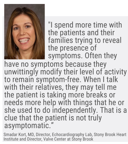
In patients with severe AS who claim to have no symptoms, “I spend more time with the patients and their families trying to reveal the presence of symptoms,” she says. “Often, they have no symptoms because they unwittingly modify their level of activity to remain symptom-free. When I talk with their relatives, they may tell me the patient is taking more breaks or needs more help with things that he or she used to do independently. That is a clue that the patient is not truly asymptomatic.”
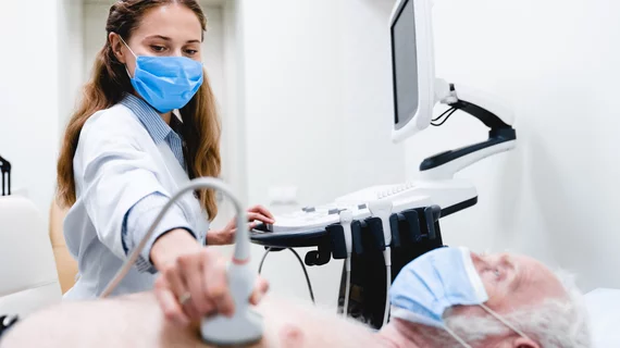
Kort performs an exercise stress echo on patients that such conversation fails to reveal the presence of symptoms. It helps by establishing their exercise tolerance in an objective way. In addition to stress- induced symptoms, she also looks for additional parameters such as changes in ECG and BP, LV systolic function using EF and global longitudinal strain as well as diastolic filling pressures. “The beauty of this test is that it is non-invasive, does not involve radiation, and is safe to do as long as the patient does not have symptoms at rest,” she notes. “It allows me to monitor the patient periodically and look for progression of their disease over time. Using the information obtained from the patient and family together with the results of the resting and stress echoes and possibly additional diagnostic modalities, an informed discussion can then take place about the next steps which may include an intervention.”
She calls this “the beauty of cardiology—combining subjective data from patients and relatives with objective scientific data for making shared decisions.”
For patients who don’t have evidence of severe AS on their resting echo, her team spends time excluding the presence of symptoms. “We rely on the technologies and tools we have to make sure we are not missing severe disease or associated pathology that could explain the symptoms,” she says. “In the echo lab, we have more sensitive parameters than EF like strain imaging to assess the systolic function of the heart. We use it a lot in patients with valvular heart disease to assess function and make sure we don’t get to a point that the ejection fraction drops and the damage is irreversible.
When indicated, Kort’s team uses TEE [transesophageal echo] to visualize the valve better and see the leaflets, how calcified they are and how well they open. “Stress echo is a fantastic tool for assessing symptomatic patients with moderate disease on resting imaging,” she says. “It allows us to assess the effect of exercise on the valve and the ventricle and to correlate stress induced findings with exertional symptoms.”
Some patients with small aortic valve areas have low gradients despite having normal flow and preserved EF. “We believe that most of them have moderate AS because as a group they have the best prognosis of all AS patients,” she notes. “However, some of them have severe AS and would not do well if left untreated. It is critical that we identify those who could benefit from intervention, and stress echo is a great tool we have in our armamentarium.”
Better detection of this low-flow, low-gradient AS patient base and potentially guiding these patients to a needed intervention comes down to careful listening across cardiologists, physicians and heart teams. Especially now during this pandemic it is important for all physicians caring for patients with valvular heart disease to check on their patients often. “Tele health is a great tool that allows us to talk to patients and their relatives without bringing them to the office,” she says. “Many patients have modified their level of activity as they hide at home and assessing for the presence of symptoms is even more challenging. We call them to ensure there has been no progression of their disease, and to remind them we are here for them had they experienced any new or worsening symptoms. We have created a safe environment for patients who do need to come in for additional tests or interventions and together with the patient we can decide on the optimal timing of that.”
References:
1. Lindroos M, Kupari M, Heikkilä J, Tilvis R. Prevalence of aortic valve abnormalities in the elderly: an echocardiographic study of a random population sample. J Am Coll Cardiol. 1993;21(5):1220–1225.
2. Otto CM, Kuusisto J, Reichenbach DD, Gown AM, O’Brien KD. Characterization of the early lesion of ‘degenerative’ valvular aortic stenosis. Histological and immunohistochemical studies. Circulation. 1994;90(2):844–853.
3. Lewin MB, Otto CM. The bicuspid aortic valve: adverse outcomes from infancy to old age. Circulation. 2005;111(7):832–834.

