New expert guidelines: Start PHV evaluations with echocardiography, but other imaging modalities can provide value
The American Society of Echocardiography (ASE) has published new recommendations on the evaluation of prosthetic heart valve (PHV) function using cardiovascular imaging. The document, made in collaboration with both the Society for Cardiovascular Magnetic Resonance (SCMR) and Society of Cardiovascular Computed Tomography (SCCT), represents an update of ASE’s original guidelines on the topic from 2009.
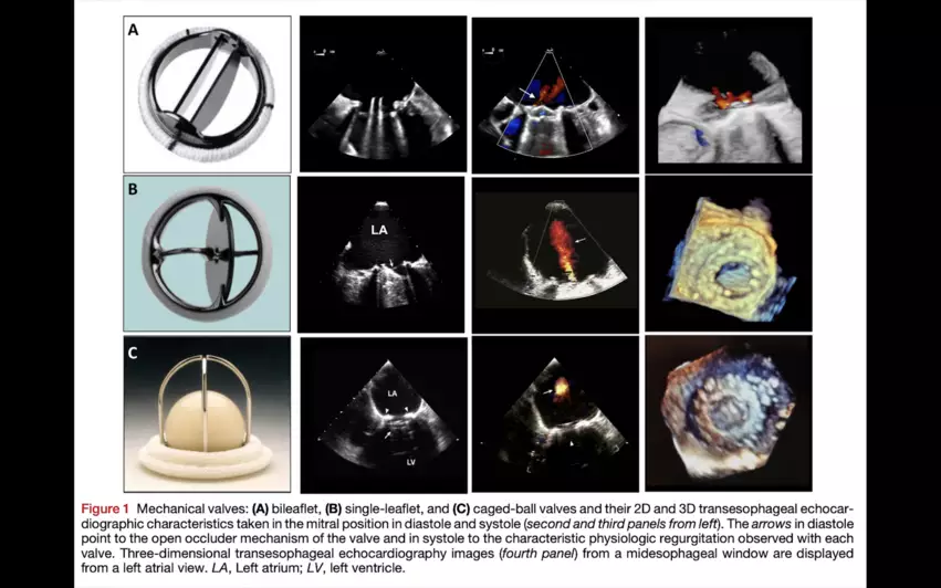
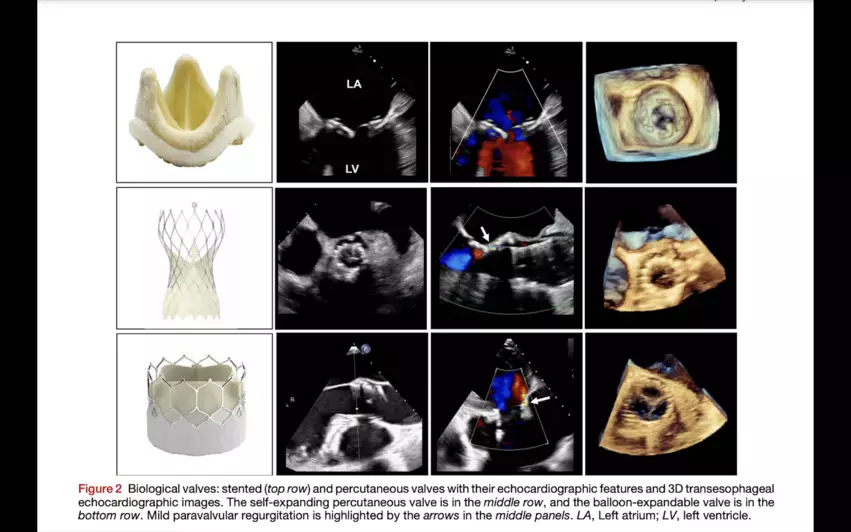
Bioprosthetic valves, including the self-expanding and balloon-expandable devices used during transcatheter aortic valve replacement (TAVR) procedures, and mechanical valves are both covered at length in the new guidelines.
Echocardiography remains the preferred imaging option for any initial evaluations, according to the new recommendations. However, recent advances in cardiac CT and cardiac MR have helped those modalities become increasingly useful when diagnosing and managing today’s heart patients. This was one of the primary reasons ASE and its collaborators wanted to update the prior recommendations from 15 years ago.
PHVs can be more challenging to evaluate than native valves, the groups added, due to “suboptimal visualization” and the “inherent variability” of a market that includes so many different valve types and sizes. The authors intend this updated document to help ease some of that confusion.
“This new guideline on prosthetic valves was very much needed, as the field has changed so much since 2009, with the introduction of percutaneous valves and improvements in 3D echocardiography and multimodality imaging,” William A. Zoghbi, MD, chair of the writing group and a chairman of the department of cardiology at Houston Methodist Hospital in Texas, said in a prepared statement. “It provides clinicians with a roadmap for evaluating PHVs, while aiming to improve patient care and outcomes in the field.”
“The new guideline provides the clinician with much-needed information on how to evaluate PHVs with cardiac ultrasound, particularly with the added value of 3D echocardiography, and when to use further imaging with cardiac CT or CMR,” added Pei-Ni Jone, MD, co-chair of the writing group and director of the echocardiography lab at Ann & Robert H. Lurie Children’s Hospital of Chicago.
The updated guidelines have been published in the Journal of the American Society of Echocardiography.[1] Click here to read the document in full.


