
Good to great: 5 ways to help sonographers deliver better echocardiograms and improve the diagnosis of severe AS
While the symptoms of severe aortic stenosis (AS) can vary patient to patient, a good echocardiogram can help a cardiologist make a quick and definitive diagnosis. But achieving consistently good echo exams is often a challenge across sonographers and facilities. Here’s some expert advice from three cardiologists on helping sonographers go from good to great on echo exams.
While severe AS affects an estimated 3.4% of Americans 75 years old and older, it remains both underdiagnosed and undertreated.[1] One way cardiologists and the rest of the heart team can reverse that trend is working to improve the quality of echocardiograms. Getting all sonographers on the same page—delivering accurate, complete examinations that give cardiologists all of the information they need—can help heart teams identify and treat more patients with severe AS than ever before.
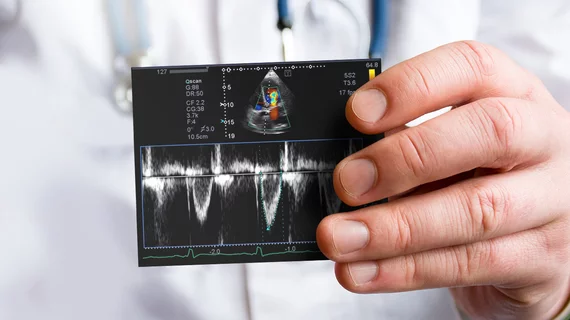
“Echocardiography, and in particular transthoracic echocardiography, is the primary means for screening and establishing the diagnosis of AS,” explains veteran cardiologist Muhamed Saric, MD, PhD, director of the Noninvasive Cardiology Lab at NYU Langone Health in New York City. “There are other imaging modalities that can serve certain roles, but nothing is more important than echocardiography. This really puts the onus on sonographers to do their jobs well day in and day out.”
Saric is one of the country’s leading experts on echocardiography and structural heart disease. While he specializes in cardiac imaging, with his team performing approximately 100,000 echocardiograms per year, he was also part of the New York University team that performed the world’s first transeptal transcatheter mitral valve replacement of a native valve. Throughout his long career, he’s seen firsthand how much cardiologists rely on their sonographers to provide high-quality images.
“Sonographers are extremely important members of the team,” he adds. “People sometimes lose sight of that fact, but it’s very true.”
So, what can hospitals and healthcare systems do to help their sonographers deliver better, more accurate echocardiograms and improve the diagnosis of severe AS? Here are five ideas.
1. Invest in your team: Make continued training and education a priority
Sonographers don’t achieve greatness overnight; it takes years of hard work, continued learning and dedication. With little to no mentorship, they are unlikely to improve as time goes on. But with continuous training and feedback, they’ll learn, grow, and reach new heights.
Helping sonographers develop also means giving them what they need—when they need it—to provide the very best care possible.
“It starts with investing in your sonographers,” says Elana Koss, MD, director of Structural Heart Echocardiography at North Shore University Hospital, Northwell Health on Long Island, New York. “That means training them, giving them high-fidelity equipment, and giving them time to do a complete and thoughtful echocardiogram.”

“It starts with investing in your sonographers. That means training them, giving them high-fidelity equipment, and giving them time to do a complete and thoughtful echocardiogram.”
- Elana Koss, MD, director of Structural Heart Echocardiography at North Shore University Hospital
Koss adds that resources such as the national guidelines, CME and videos developed by the American Society of Echocardiography are a great way for sonographers to keep up with a field that is ever-changing, but communication, direct feedback, hands-on-training and cultivating a team approach are ultimately most impactful to helping sonographers realize their full potential.
“Teaching your sonographers, investing in their development and creating a consistent feedback loop, will elevate the quality of their scanning and improve diagnostic accuracy. This benefits the patient which is what this is all about,” she says. “Sonographers are eager for that level of engagement and impact. They want to be involved and know that what they are doing matters for patient care.”
Saric agrees and is quick to emphasize that cardiologists, sonographers and everyone else involved in the diagnosis of severe AS already have the tools they need to be great at their jobs.
“First and foremost, we have to rely on standard practices that have been established since the 1980s,” he says. “Any shortcomings in recognizing AS do not come from a lack of new technologies or unprepared labs; it’s all about learning and honing those established techniques.”
Saric often travels around the United States to discuss best practices with sonographers. During these gatherings, they bounce from one key topic to the next such as the importance of the Dimensionless Index or how to recognize calcification in the valve. Providing continuous education, he says, is another way to help sonographers fine-tune their skills and improve their chances of performing a successful exam.
Saric believes in teaching sonographers as much as possible about the care process. If they can gain a better understanding of interventional procedures such as transcatheter aortic valve replacement and more invasive treatments such as surgical aortic valve replacement, it provides context that they can carry with them going forward.
“Understanding these different procedures can really help sonographers understand why they do what they are doing,” he says. “They benefit, instead of feeling like they are just performing exams and making these abstract measurements, when they are given additional information about why patients need to be treated and how failing to treat AS can lead to higher mortality rates and even sudden death.”
2. Make sure sonographers practice, practice and then practice some more
Practice may not necessarily make perfect, but it can go a long way toward helping sonographers deliver the level of work that cardiologists and other physicians expect. Koss recommends that sonographers take advantage of every opportunity to perform a complete echocardiogram. More than 60% of patients with aortic stenosis have their highest gradients from non-apical views.2 Not exploring these views can lead to misdiagnosis and misclassification of aortic stenosis in a significant number of patients. That is humbling. Even if the patient isn’t yet at the point of significant AS, making a habit of acquiring these views—a right parasternal view or a suprasternal notch view, using the Pedoff non-imaging probe—hones those skills over time. Learn from your colleagues and seek assistance from a more experienced sonographer if needed. These are some different ways to help ensure accuracy when a patient with AS is being evaluated.
“At the end of the day, the most important thing is to practice,” she says. “Seek feedback, implement what you have learned, practice it and repeat. I tell the sonographers that they are the front line for diagnosis of AS—we can’t treat what we don’t diagnose and we can’t accurately diagnose what we don’t see.”
3. Tell sonographers exactly what you need—and give them enough time to deliver
Providing sonographers with clear, reasonable expectations is a quick way to get everyone on the same page. For example, Koss says cardiologists will often encounter echocardiograms that only include a single view, but a minimum of two views—and ideally even more than that—are needed to make an accurate diagnosis. When a sonographer identifies some degree of AS, they should investigate further, exploring non-apical views(i.e. right parasternal, subcostal and suprasternal notch).
Communicating and setting these expectations at the outset helps set the sonographer up for success, ensuring they capture the appropriate views the first time around, without having to bring the patient back for a repeat exam.
“Without a complete study, we are likely going to need to repeat the echo,” Koss says. “Repeat imaging is a drain on all—financially burdensome to the system and the patient, and a waste of the sonographer’s, cardiologists, and patient’s time. The goal should always be to never repeat a study.”
Of course, sonographers can only do so much with the time they are given. If you rush them—giving them only 20 minutes per exam, for example—they are less likely to do a thorough job. Imaging a young, healthy patient might take 20 minutes, but a very sick, complex patient with multi-valve disease can take 45 minutes to an hour to scan, even for an experienced sonographer.
“If you expect a lot from your sonographers, you need to give them the tools and support they need to succeed,” Koss says.
4. Highlight the positive
Most institutions responsible for providing echocardiography services have monthly or even bi-weekly quality control (QC) meetings, and larger training hospitals typically meet weekly to review the work of trainees and fellows. Saric, who hosts weekly QC meetings at NYU Langone, says these regular gatherings represent a perfect opportunity to highlight the importance of sonographers in front of the rest of the care team.
“I always make it clear that none of the exams we reviewed would have even been performed if not for the hard work of our sonographers,” he says. “It’s important to provide that public recognition in front of a large audience when you have the chance. Some of the people in these meetings may not even realize where the reports come from or how much effort it takes.”

“When a lab submits studies performed by its sonographers or interpreted by its physicians, one of the main things being looked at is how well aortic stenosis was evaluated. So in addition to being important from a medical point of a view, aortic stenosis is also a key element when trying to demonstrate the quality of care you can provide."
- Muhamed Saric, MD, PhD, director of the Noninvasive Cardiology Lab at NYU Langone Health
5. Push for lab accreditation
Organizations such as the Intersocietal Accreditation Commission (IAC) offer accreditation services that help echocardiography labs demonstrate their high quality standards to patients and other healthcare providers. Receiving accreditation is a signal that your lab meets all applicable requirements related to equipment, medical personnel and quality assurance.
While all accreditation guidelines are different, the IAC prioritize a lab’s ability to identify and diagnose AS. Any facilities seeking accreditation will need to submit a completed echocardiogram that shows clear signs of AS.
“These certifying bodies have decided that identifying AS is a key parameter to a lab’s success,” Saric says. “When a lab submits studies performed by its sonographers or interpreted by its physicians, one of the main things being looked at is how well AS was evaluated. So in addition to being important from a medical point of a view, AS is also a key element when trying to demonstrate the quality of care you can provide. I think we should encourage every echocardiography lab in the United States to seek accreditation.”
Click here for additional information on evaluating AS.

Michael has more than 19 years of experience as a professional writer and editor. He has written at length about cardiology, radiology, artificial intelligence and other key healthcare topics.