Magnetic Resonance Imaging

Magnetic resonance imaging (MRI) is the gold standard imaging modality for soft tissues. It produces detail cross-sectional images of soft tissue and bone anatomy, including muscles, tendons, ligaments, brain and organs, without the use of ionizing radiation. In addition to orthopedic imaging, MRI is also used for heart, brain and breast. MRI uses gadolinium contrast in many exams to highlight tissues and blood vessels, which enhances images and offers better diagnostic quality. It can also be used in conjunction with PET scans. How does MRI work? MR creates images by using powerful magnets to polarize hydrogen atoms in water (the body is made of of more than 80% water) so they face in one direction. A radiofrequency pulse is then used to ping these atoms, causing them to wobble, or resonate. The MRI coils detect this and computers can assemble images from the signals. Basic MRI scans will focus on the resonance of fat and water in two different sequences, which highlight and contrast different features in the anatomy.

Cardiac PET on the rise among U.S. cardiologists

SPECT is still the most common modality used to evaluate CAD patients, but cardiac PET is gaining more and more momentum.

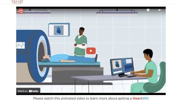
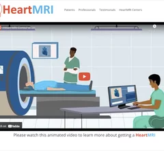
The Society of Cardiovascular Magnetic Resonance (SCMR) recently launched HeartMRI.org, which provides includes a general information and videos for patients and referring physicians.

SCMR launches cardiac MRI educational video to assist referring physicians 

The Society of Cardiovascular Magnetic Resonance launched a new website aimed at patients and referring physicians that includes pamphlets and videos on cardiac MRI. 

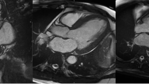
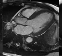
CMR of a patient showing evidence of myocardial hypertrophy

MRI results help cardiologists predict heart failure risk

"This breakthrough suggests that heart MRI could potentially replace invasive diagnostic tests," one specialist said. 

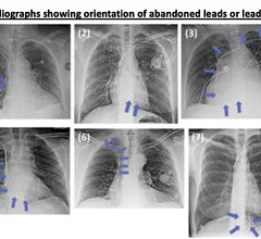
Some of the abandon leads and various shapes and positions on the wires from the study. None of these patients had any safety issues related the abandoned leads. Image courtesy of RSNA

MRI in patients with abandoned pacemaker and ICD leads

Abandoned metal wire leads for pacemakers and ICDs did not present a serious safety issue in a study of 80 patients, but a handful of patients did experience a significant change in the composite of capture threshold, sensing or lead impedance.

Medicare drops AUC requirement for advanced imaging, ASNC celebrates

"This was an unneeded burden, which was solely adding to the administrative hassles of medicine," said American Society of Nuclear Cardiology President Larry Phillips.

Kate Hanneman, MD, University Of Toronto, explains why vendors and hospitals are increasingly discussing lowing their carbon footprint by starting with radiology. 

What does radiology have to do with climate change?

Kate Hanneman, MD, explains why many vendors and hospitals want to lower radiology's impact on the environment. "Taking steps to reduce the carbon footprint in healthcare isn’t just an opportunity," she said. "It’s also a responsibility."

An example of cardiac MRI myocardial blood flow reserve (MBFR) imaging. Image courtesy of https://doi.org/10.3390/diagnostics10090679

Imaging group thrilled with new CPT codes for MBFR measurements

The Society for Cardiovascular Magnetic Resonance applauded two new CPT Category III codes for myocardial blood flow reserve (MBFR) assessments.

Thumbnail

Multidisciplinary quality improvement initiative significantly improves on-time starts for cardiac MRI

Such delays can result in longer patient fasts, extended wait times, and poor synchronization of anesthesia induction, researchers detailed in JACR

Around the web

Ron Blankstein, MD, professor of radiology, Harvard Medical School, explains the use of artificial intelligence to detect heart disease in non-cardiac CT exams.

Eleven medical societies have signed on to a consensus statement aimed at standardizing imaging for suspected cardiovascular infections.

Trimed Popup
Trimed Popup