
Better than BMI? 3D body scanner uses AI to measure metabolic health
Researchers have found that an unexpected combination—artificial intelligence (AI) and a 3D body scanner originally used to design clothing—can evaluate a person’s metabolic health and identify significant risks of cardiovascular disease, diabetes and other adverse outcomes. In fact, the technique may prove to be more accurate than knowing a person’s body mass index (BMI) or waist-to-hip ratio.
A team of researchers at Mayo Clinic in Rochester, Minnesota, made the discovery, sharing their findings in European Heart Journal - Digital Health.[1]
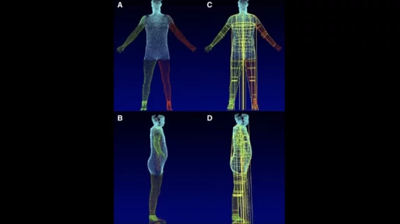
The group started by training and validating an advanced AI model with data from nearly 1,300 individuals who volunteered to go inside a 3D body-volume scanner and then undergo a series of tests and measurements. The scanner in question was originally used to help design and size pieces of clothing.
An additional 133 volunteers were brought in to provide front- and side-view images taken with the myBVI mobile phone app to further explore the new technique’s potential.
Overall, the AI-powered body scanner was confirmed to be more accurate at identifying individuals who may face a higher risk of metabolic syndrome than BMI or waist-to-hip ratio. When validating the technique on images captured with the myBVI mobile app, it delivered an “excellent performance.”
“There is a need for a reliable, repeatable measure of metabolic syndrome risk and severity,” first author Betsy Medina Inojosa, MD, a research fellow at Mayo Clinic, said in a statement. “BMI measurements and bioimpedance scales that measure body fat and muscle are inaccurate for many people, and other types of scans are not widely available. Our research shows that this AI model may also be a tool to guide clinicians and patients to take action and seek outcomes that are a better fit for their metabolic health.”
“This small study finds that digitally measuring a patient's body volume index with 3D imaging provides a highly accurate measurement of shapes and volumes in critical regions where unhealthy visceral fat is deposited, such as the abdomen and chest,” added senior author Francisco Lopez-Jimenez, MD, Mayo Clinic’s director of preventive cardiology. “The scans also record the volume of hips, buttocks and legs—a measure related to muscle mass and 'healthy' fat. The 3D information about body volume in these key regions, whether from the large, stationary 3D scanner or from the mobile app, accurately flagged the presence and severity of metabolic syndrome using imaging instead of invasive tests.”
Click here to read the full analysis in European Heart Journal – Digital Health, a publication from the European Society of Cardiology.

Michael has more than 19 years of experience as a professional writer and editor. He has written at length about cardiology, radiology, artificial intelligence and other key healthcare topics.