
Cardiology now has more than 100 FDA cleared AI algorithms
Cardiology is ranked second for the most clinical artificial intelligence (AI) algorithms cleared by the U.S. Food and Drug Administration based on the FDA's latest update on approvals released in November.
There are 71 AI algorithms listed under the category of cardiology and another 31 cardiology specific imaging algorithms listed under radiology, bringing the total number of cardiac apps up to 102. Cardiology makes up 10% of the 692 market-cleared clinical AI algorithms as of July 30. Additional clinical AI algorithms have also been cleared since July, but were not included in an FDA update.
Radiology ranked No. 1 with 521 AI FDA approvals and accounting for 76% of all the FDA-cleared algorithms. Cardiology is a distant second, followed, by neurology with 16 algorithms in third. Many applications on the list for other specialities outside of radiology are directly related to image post-processing, so medical imaging overall accounts for more than 80% of the FDA cleared algorithms.
AI has been a key trend at all cardiology meetings in recent years. While a lot of this has been focused on research, there is also a lot of discussion in sessions on actual clinical implementation and on the expo floors of conferences where this commercialized AI is being demonstrated.
"What I want to make sure people understand is that although AI is going to largely replace what we are currently doing today, we must assure that human intuition remains at the helm of clinical decision-making and to avoid the assumptions that the machines are always right," explained Steven Lester, MD, a consultant with the department of cardiology at Mayo Clinic College of Medicine and Science, medical director of Discovery Oasis, and founder and chief medical officer of the Mayo Clinic and ASU MedTech Accelerator. He spoke on AI at the American Society of Echocardiography (ASE) 2023 meeting.
"Something truly transformational is happening with AI. We are already seeing how AI is reshaping our modern lives, transforming industries, and it is going to impact probably every aspect of healthcare," Lester said.
AI is already used every day in our lives in the form or weather apps, Google Maps, search engines, aircraft navigation, social media voice commands for devices, and many other areas that may not be obvious. That same transformation is also taking place in cardiology with automated identification and segmentation of anatomy and the AI automatically performing measurements based on the ideal, text-book views. This is leading to measurements that are more reproducible and accurate than even the best human imagers can do consistently, he said.
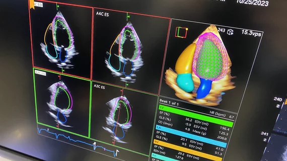
Deep learning and generative AI will fundamentally change echocardiography
The introduction of deep learning and generative pre-training transformer (GPT) algorithms are two major inflection points in the development of AI in cardiac ultrasound that will lead to major changes in echocardiography workflows in the near future. This will fundamentally change how cardiologists and sonographers do their jobs, explained cardiology AI expert Partho Sengupta, MD, DM, Henry Rutgers Professor of Cardiology and chief of cardiovascular medicine at Robert Wood Johnson Medical School and chief of the cardiovascular service line at Robert Wood Johnson University Hospital.
"Deep learning is going to completely automate all the measurements and these will auto populate on the report. This is real and it is coming. Some of this is going to be built into the ultrasound machines, and it is really going to offload the technologists and doctors who feel so burned out doing all these measurements," Sengupta explained. "In the next two or three years, everything in echocardiography will be automated with AI in terms of measurements."
These predictions are being echoed by many echocardiography experts.
"The first thing I will tell you is that in the near future, echo labs that do not have AI will fall way behind. AI is something that will live inside an echo machine. As soon as a study is done, it will be analyzed by the AI and the reader will get all the measurements. There are already many companies that are very close to having this, and I think it will change echo dramatically," explained Roberto Lang, MD, director of cardiovascular imaging at the University of Chicago and a past ASE president. "This has been happening slowly, but there will be an explosion in the next couple of years where everybody will be using AI. I don't think you can give yourself the opportunity to have a high-volume lab and not use AI. You are going to have misdiagnoses and you are not going to be very reliable on the measurements.”
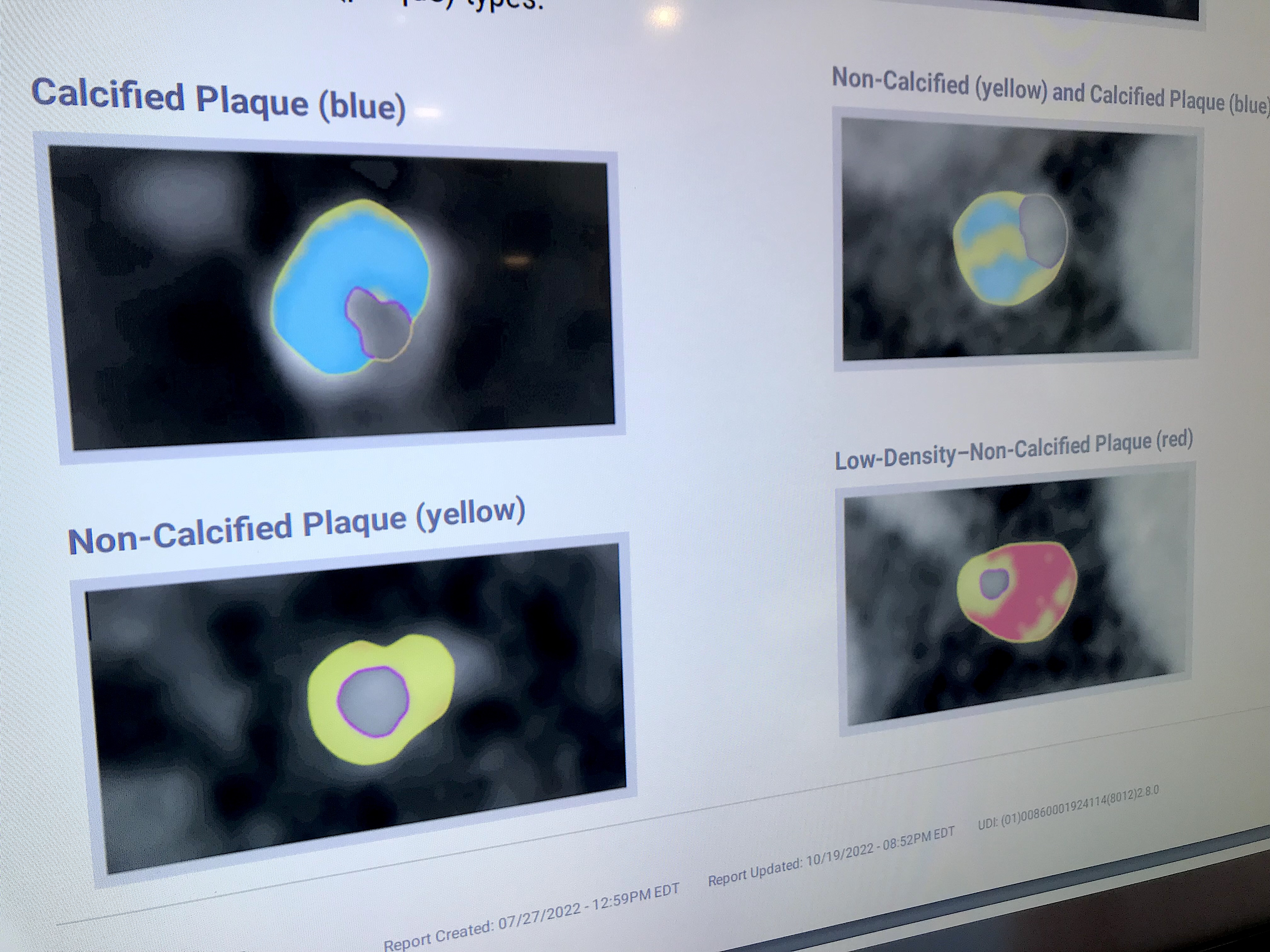
Example of an automated artificial intelligence (AI) assessment of soft coronary plaque from a CT scan from the vendor Cleerly. This image shows the different types of soft plaques the system can identify. The AI automatically identifies the plaque types, contours them and then calculates a detailed assessment with numerous measurements. This technology may be used to screen patients for early disease and then track progress made over time using various drugs to stabilize or reverse coronary disease years before it becomes symptomatic.
AI is already monitoring cardiac patients
Numerous AI clearances are for automated cardiac ECG monitoring on both hospital grade systems and consumer grade wearable watches and mobile app devices. The proliferation AI in this area is changing electrophysiology (EP).
"I think AI has a phenomenal role in not just helping with diagnosing, but with predicting and preventing disease. It also is assisting with the workflow and how we actually manage our patients," explained Jagmeet Singh, MD, professor of medicine at Harvard Medical School and founding director of the Resynchronization and Advanced Cardiac Therapeutics Program at Mass General Hospital, who spoke with Cardiovascular Business at Heart Rhythm 2023.
Singh’s primary AI research has been focused on its ability to detect atrial fibrillation (AFib) and ventricular tachycardia (VT). He has collaborated with physicians in France on a of couple studies using the Cardiologs Holter ECG analysis algorithm to analyze data from the first 24 hours of using a Holter to predict diagnoses over the next 14 days. These studies culminated in two late-breaking analyses presented at HRS the past two years.
On the expo floor of HRS, numerous vendors like Philips Healthcare, iRhythm, and Medtronic offered FDA-cleared ECG analysis algorithms for remote monitoring systems, wearable and implantable cardiovascular monitors.
Cardiac MRI and CT is being changed by AI
There are many FDA-cleared AI specific to cardiac CT and MRI. One example of it being implemented is at Brigham and Women's Hospital rolling out an AI-driven approach to cardiac MRI. The AI model picks the views and enables the acquisition with just one click. It also provides all measurements and quantification automatically.
"What that has done for us is impact many important areas. Number 1, it shortened the examinations. It is good for patients because they don't need to spend as much time in the magnet, but it is also better for throughput because we can accommodate more patients on any given day. Number 2, it has increased the quality of what we do, because it is less dependent on the expertise of the technologists who are pressing the buttons and making decisions. And then on the post-processing it has simplified our lives. What used to be a very time-consuming process is now just a click away. For me, that is very impactful," Marcelo DiCarli, MD, chief of the division of nuclear medicine and molecular imaging and executive director for the cardiovascular imaging program at Brigham and Women's Hospital, said in and interview with Cardiovascular Business.
While there is usually no reimbursement for the use of AI, the ROI is generally seen in the form of improved efficiency, workflow and accuracy in measurements.
"When it comes down to business, it is about throughput on the scanner. As reimbursement comes down, being able to replace the shrinkage of reimbursements with being able to accommodate more patients becomes very relevant. And for physician time, it also becomes very relevant. Doing our work more more efficiently, accurately and potentially even better becomes very, very meaningful," DiCarli said.
Many imagers feared AI would replace them, but it is actually enabling them to do more work with better clinical information. “I can only think of positive things coming out of AI over the next five years," DiCarli said.

An example of HeartFlow's FFR-CT technology showing the coronary tree of a patient with a color-coded overlay showing critical drops in coronary blood flow. It takes the cardiac CT dataset and uses computational fluid dynamics to create virtual fraction flow reserve values. Photo by Dave Fornell
First cardiac AI has already entered patient care guidelines
Many cardiac imaging experts say AI is no longer a science project and it is impacting real, everyday patient outcomes now. The realization that AI has arrived in the real world hit home for Society of Cardiovascular Computed Tomography (SCCT) President Ed Nicol, MD, consultant cardiologist and honorary senior clinical lecturer with Kings College London, when the first cardiac AI algorithm was included in practice guidelines in both Europe and the United States. The ACC/AHA 2021 Chest Pain Guidelines include the recommended use of AI-driven fractional flow reserve hemodynamic flow measurements derived from noninvasive CT imaging (FFR-CT).
"If you had said to me 15 years ago we were going to have some sort of AI computational fluid dynamics tool in the U.S. chest pain guidelines, you would have been laughed at. Everyone would have said you were crazy. But that is the reality, we see FFR-CT in the international guidelines, based on evidence. That will be the first, I suspect, of many," Nicol said.

Dave Fornell has covered healthcare for more than 17 years, with a focus in cardiology and radiology. Fornell is a 5-time winner of a Jesse H. Neal Award, the most prestigious editorial honors in the field of specialized journalism. The wins included best technical content, best use of social media and best COVID-19 coverage. Fornell was also a three-time Neal finalist for best range of work by a single author. He produces more than 100 editorial videos each year, most of them interviews with key opinion leaders in medicine. He also writes technical articles, covers key trends, conducts video hospital site visits, and is very involved with social media. E-mail: dfornell@innovatehealthcare.com