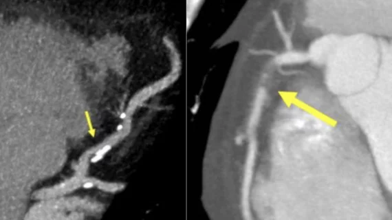
New CAD-RADS 2.0 reporting for coronary CTA offers patient management recommendations
A new expert consensus document "2022 Coronary Artery Disease – Reporting and Data System (CAD-RADS 2.0)" was released this week that expands on the first version CAD-RADS released in 2016. The original document helped standardize radiology reporting on coronary CT angiography (CCTA), and the new 2.0 version can be used to guide possible next steps in patient management.
The Society of Cardiovascular Computed Tomography (SCCT) released the new expert consensus document on CAD-RADS, in collaboration with the American College of Cardiology (ACC), the American College of Radiology (ACR) and the North America Society of Cardiovascular Imaging (NASCI).
The collaboration was led by Ricardo Cury, MD, MBA, FACC, FACR, FAHA, MSCCT, director of cardiac imaging, Baptist Health South Florida, and Ron Blankstein, MD, FACC, MSCCT, FASPC, associate director, cardiovascular imaging program, Brigham and Women's Hospital. They said the goal was to update and improve the initial 2016 reporting system for CCTA by incorporating the latest technical developments as well as recent clinical trials and guidelines in cardiac computed tomography (CCT).
“Even though coronary CTA can be a very useful test when performed on the right patient population, the test itself does not change outcomes," explained Blankstein in a statement from SCCT. "Rather, it is how clinicians act on the test results that ultimately makes a difference. For this reason, it is essential to provide referring clinicians with patient management recommendations which are now part of the CAD-RADS 2.0 statement."
Cury helped write the first set of CAD-RAD guidelines in 2016, which were designed enable apples-to-apples comparisons between patients and in serial exams and attempted to standardize taxonomy aused in reports and remove some of the variability between radiologists and cardiologists reading the studies.
"The new guidelines and increasing data supporting the prognostic relevance of coronary plaque burden and physiologic assessment by cardiac CT," the SCCT statement read.
Cury said the document considers the newest data recent clinical trials. He stressed that, because of this update, CAD-RADS will continue to provide a framework of standardization that may benefit education, research, peer-review, artificial intelligence development, clinical trial design, population health and quality assurance with the ultimate goal of improving patient care.
The document was published in the Journal of Cardiovascular Computed Tomography (JCCT).[1]
What is new in CAD-RADS 2.0?
It includes updated classification to established a framework for stenosis, plaque burden and plaque modifiers, which will include assessment of CT fractional-flow-reserve (CT-FFR) or myocardial CT perfusion (CTP), when performed.
SCCT said a key update is that plaque burden should be estimated whenever present. This can be accomplished using either an evaluation of the amount of coronary artery calcium (CAC), segment involvement score (SIS), a visual assessment, or total plaque burden quantification, when available and validated. Based on these methods, the overall amount of plaque (P) descriptor ranges from P1 to P4 (mild, moderate, severe, extensive) to denote increasing categories of plaque burden.
Two new modifiers were also added. Modifier I (ischemia) indicates that an ischemia test with CT has been performed (either CT-FFR or stress CTP) and if the result was positive, negative or borderline. The second modifier E (exceptions) was added to denote the presence of non-atherosclerotic narrowing of the coronary arteries.
Along with significant updates, some consistencies remain from the original guideline and were emphasized in the 2.0 version, including the stenosis severity classification remains the same, ranging from CAD-RADS 0 for absence of any plaque or stenosis to CAD-RADS 5 indicating the presence of at least one 100% occluded vessel.
The authors also stressed that CAD-RADS classification should always be interpreted together with the impression found in the report.
Related CAD-RADS Content:
Radiologists utilize novel CAD-RADS in 95% of coronary CTA reports
CAD-RADS a ‘big step in the right direction’ toward improving outcomes for acute chest pain
Plaque characteristics boost predictive power of CTA risk scoring
Continued variation in radiology tech reports poses threat to readability
Find more cardiac CT news and video
Reference:

