
PHOTO GALLERY: Cardiac CT advances at SCCT 2022
This photo gallery includes images from the 2022 Society of Cardiovascular Computed Tomography (SCCT) meeting, which took place July 14-17 in Las Vegas. The meeting was the largest for SCCT since the society was founded, partly driven by the increasing interest in cardiac computed tomography angiography (CTA), which now has a 1A recommendation for front line imaging in the ACC 2021 Chest Pain Guidelines.
Several vendors showed new technologies, including artificial intelligence (AI)-driven software to assess soft coronary plaques. One of the main takeaways from the meeting was this technology may offer a paradigm shift in cardiology with very early detection of disease years prior to symptoms, enabling true preventive care.
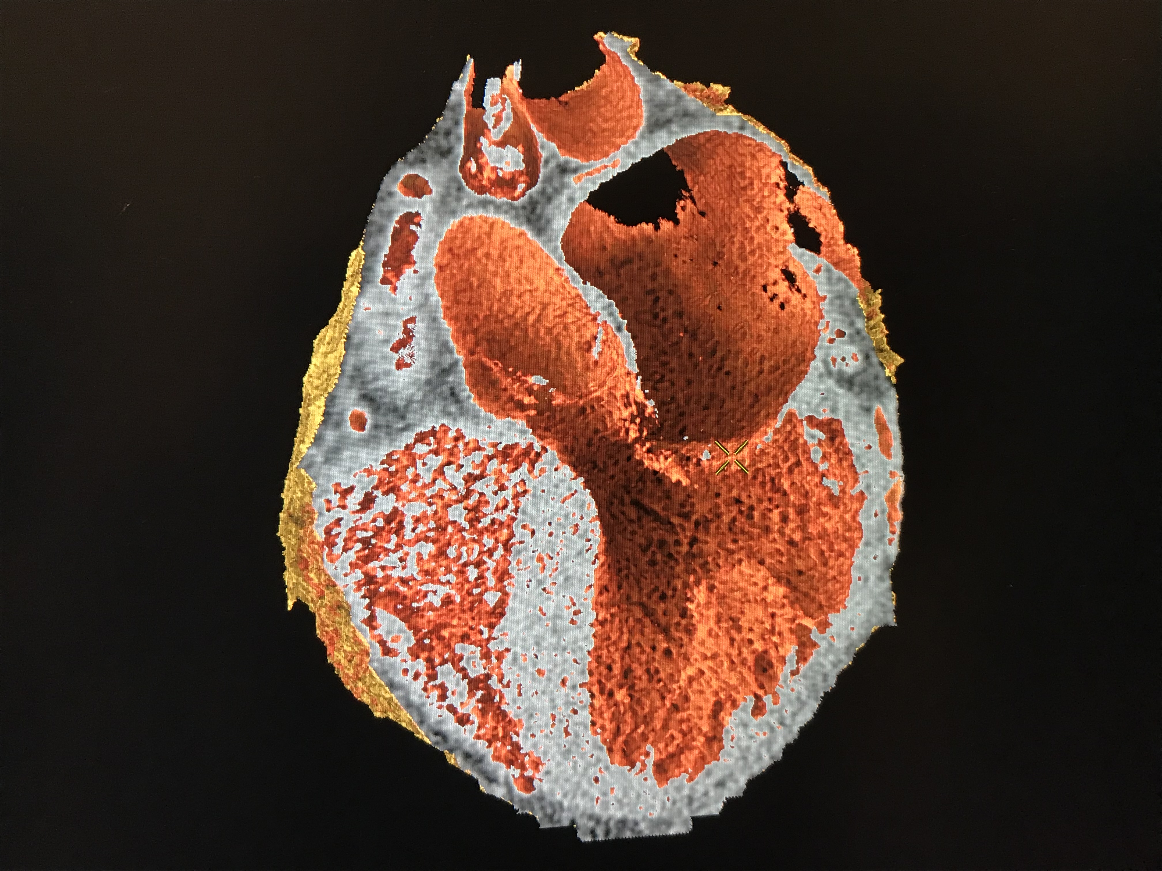
Example of photo-realistic CT imaging using the GE Healthcare Volume Illumination feature on its post-processing software, demonstrated on the expo floor of SCCT.
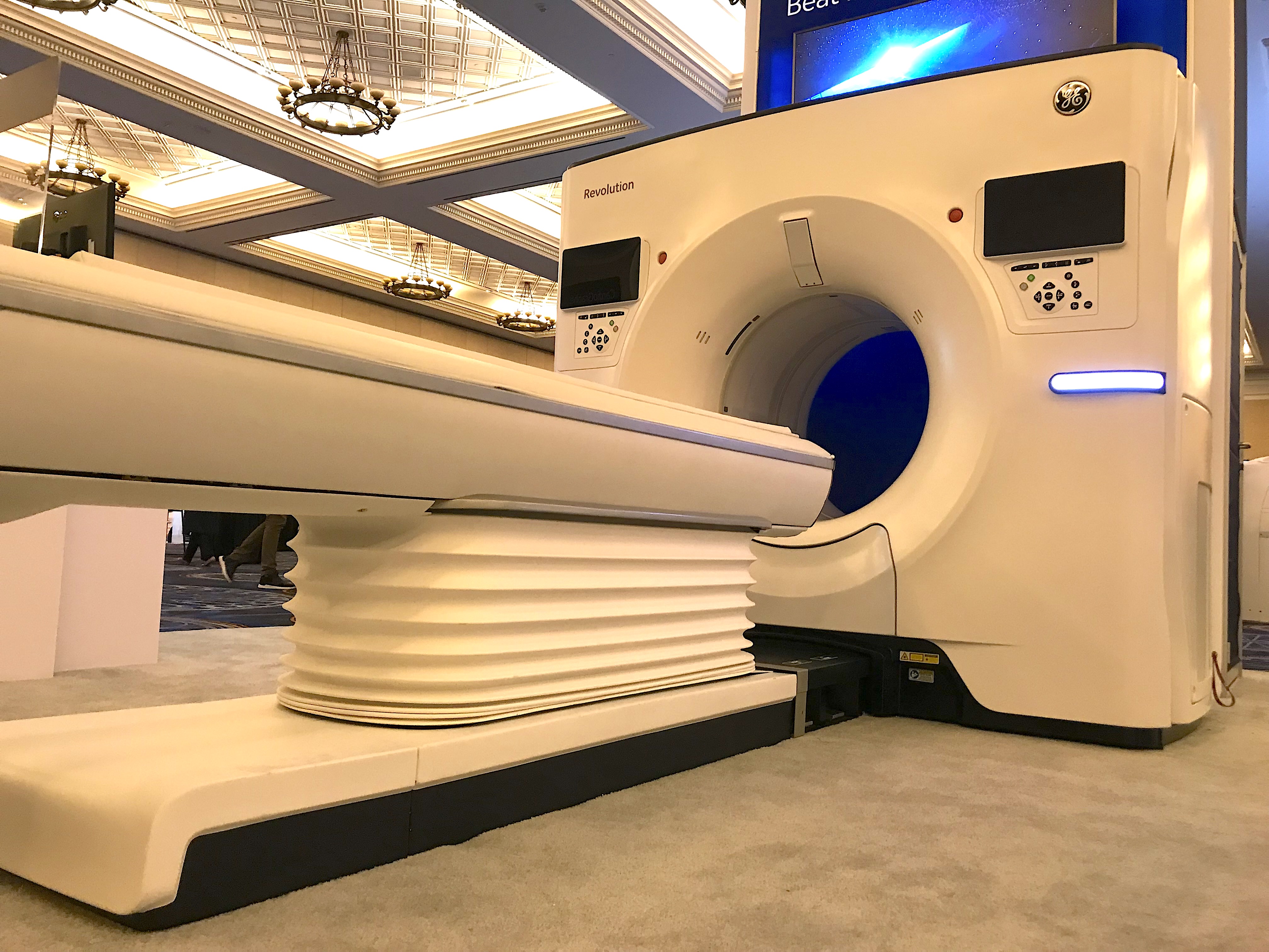
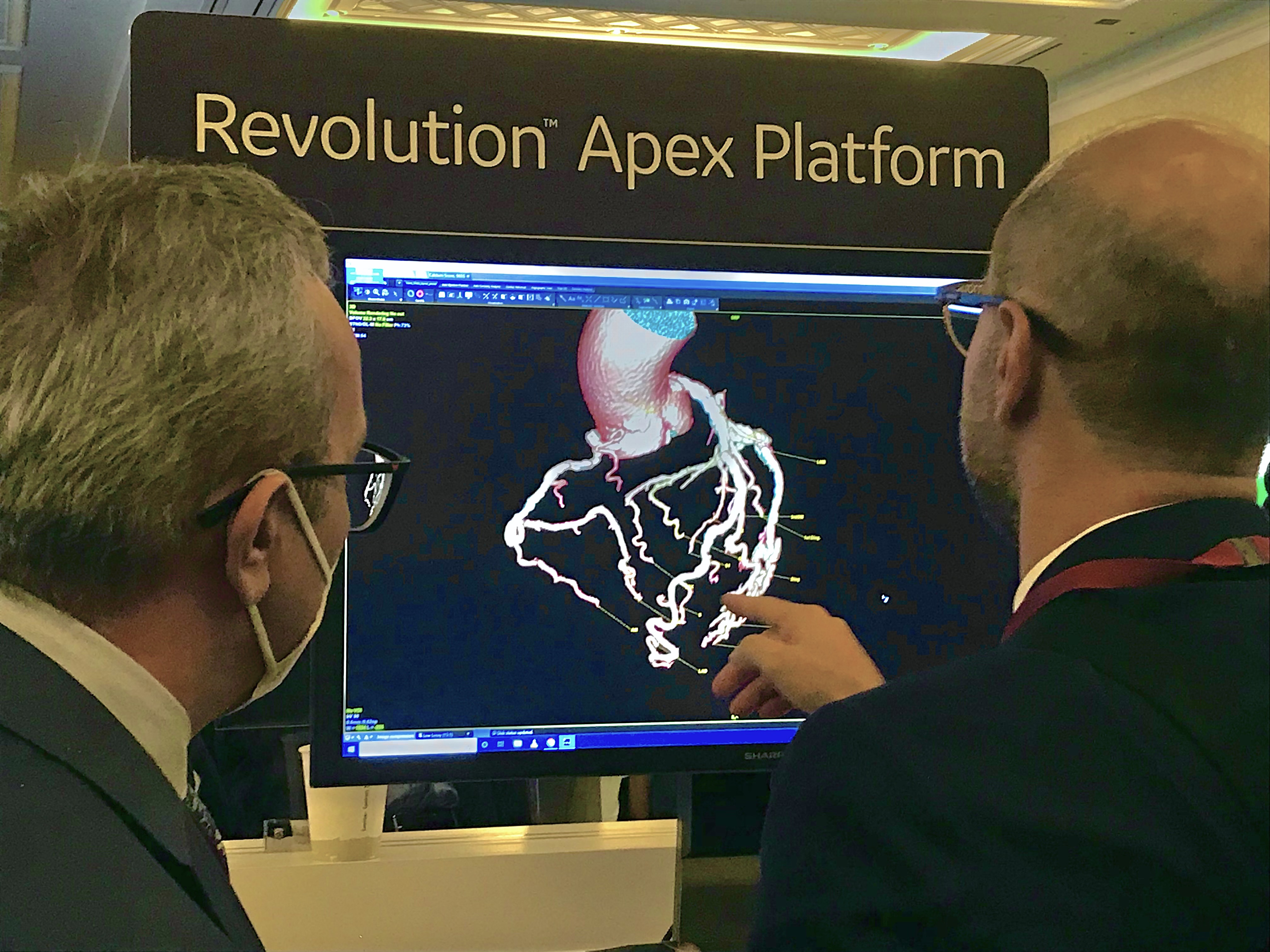
GE displayed its new Revolution Apex CT system optimized for cardiac scanning at the 2022 Society of Cardiovascular Computed Tomography (SCCT) meeting. The system also can be used as a general radiology scanner. GE is among the vendors that are really promoting CTA in the wake of the 2021 chest pain guidelines that elevated CTA to a 1A recommendation.
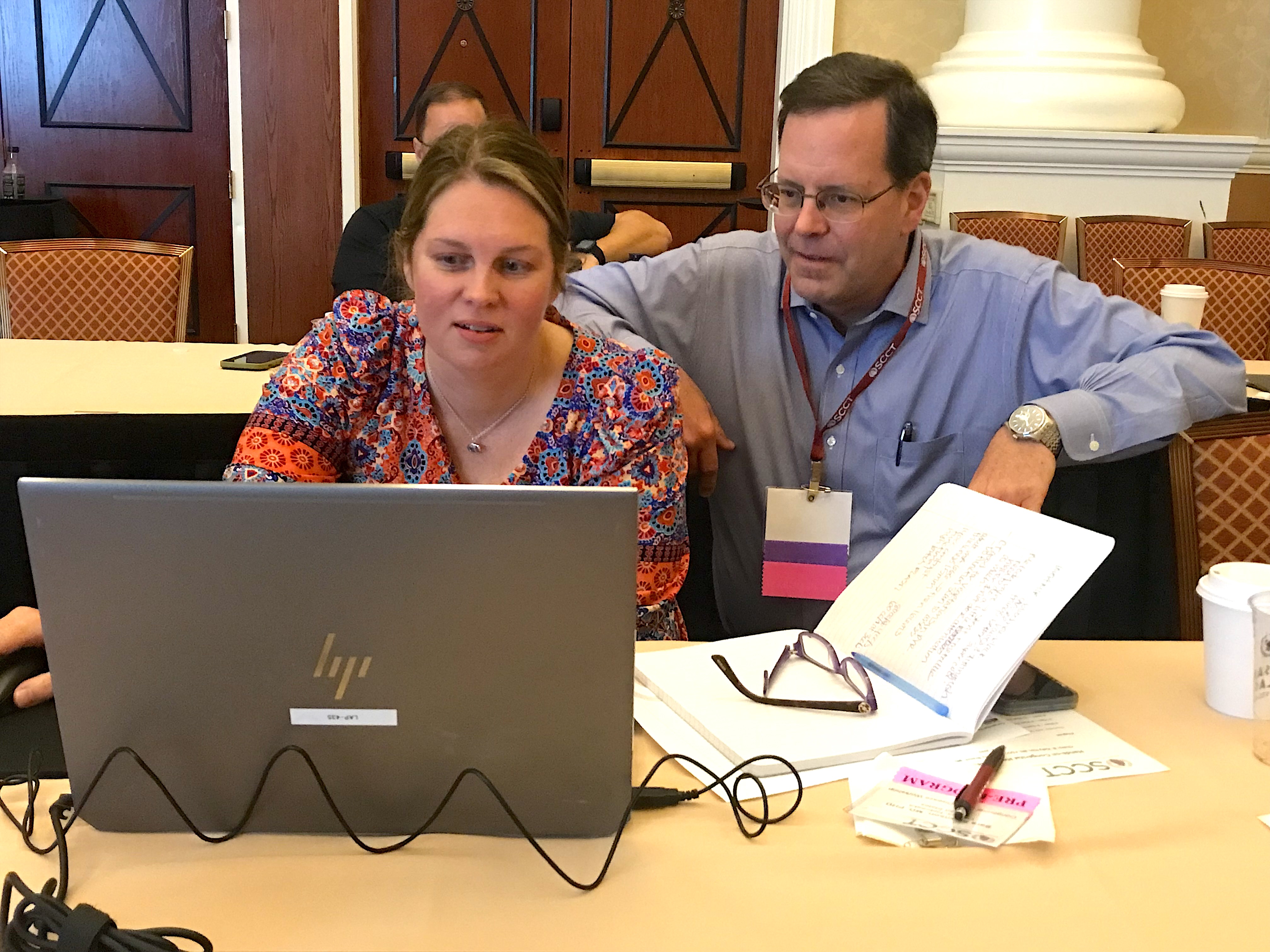
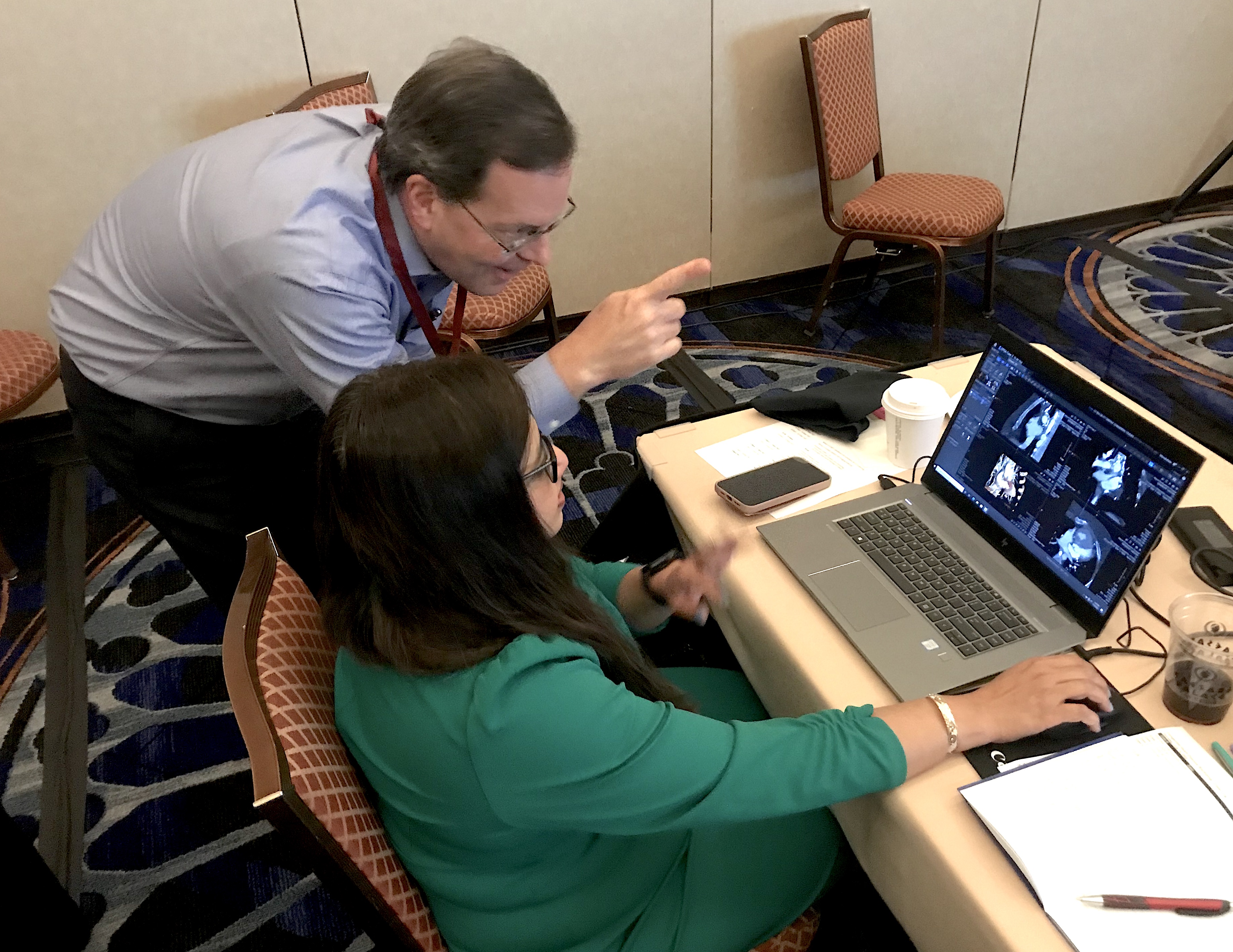
Pre-conference all-day training course for the Coronary CTA Academy: Cases from Basic to Intermediate. CTA experts explained step by step how to review cardiac CT scans and how to do basic post-processing of the datasets.
Left, coronary CT angiography of a vessel showing plaque heavy calcium burden. Right, image showing color code of various types of plaque morphology showing the complexity of these lesions that was automatically created using artificial intelligence. Cardiac AI vendor Elucid has developed the now FDA-cleared algorithm to automatically assess a coronary CT scan to identify all the plaque in the vessels, classify it and create a report with images. It is widely believed by SCCT experts this type of technology may change the direction of cardiology toward more preventive care and offers the ability to monitor plaques. Read more on this trend in the article Cardiac CT soft plaque assessment my offer paradigm shift for coronary disease screening.
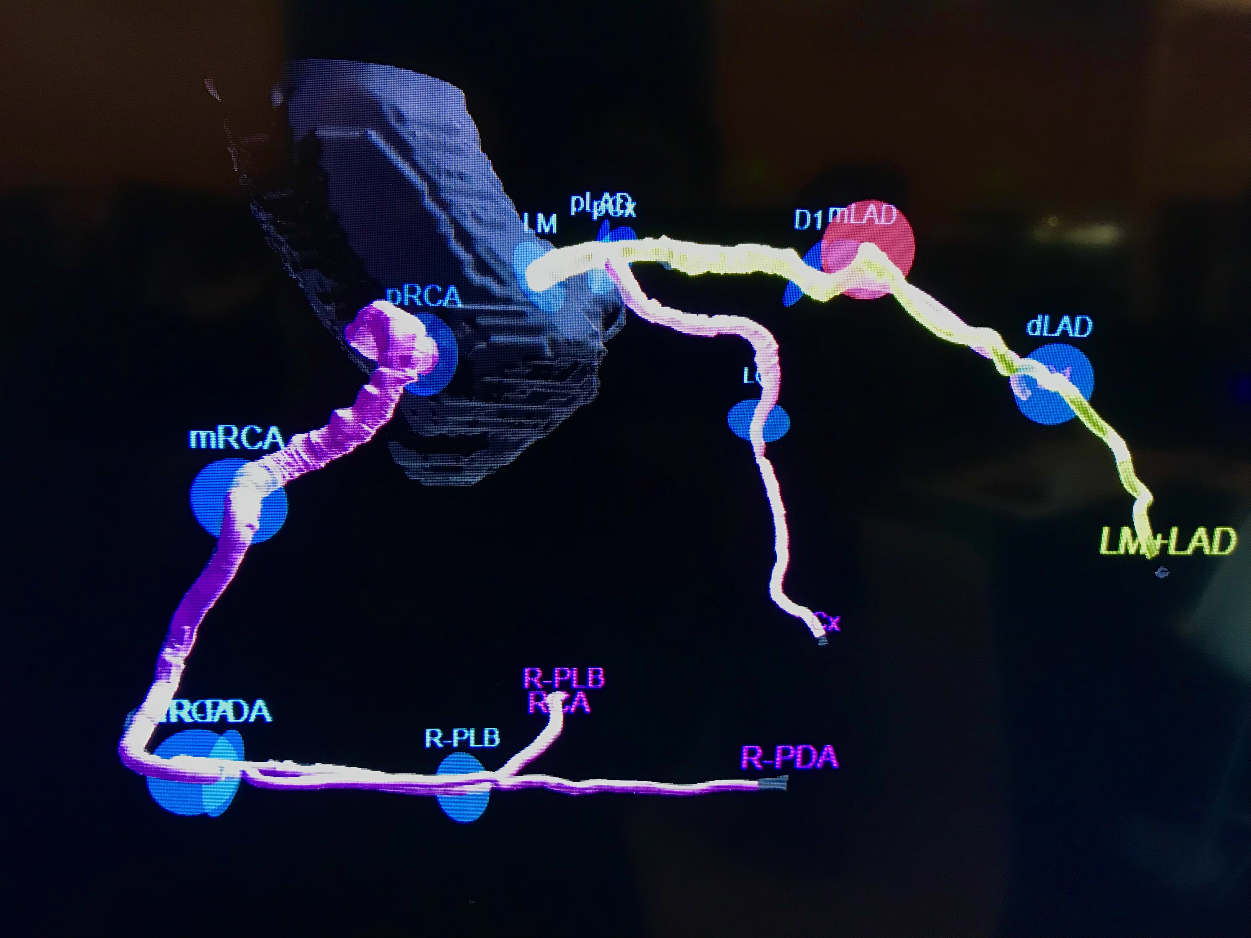
Example of a curved multiplaner reconstruction (Curved MPR) image showing the entire coronary artery in one frame for easier evaluation of the plaques and lumen.
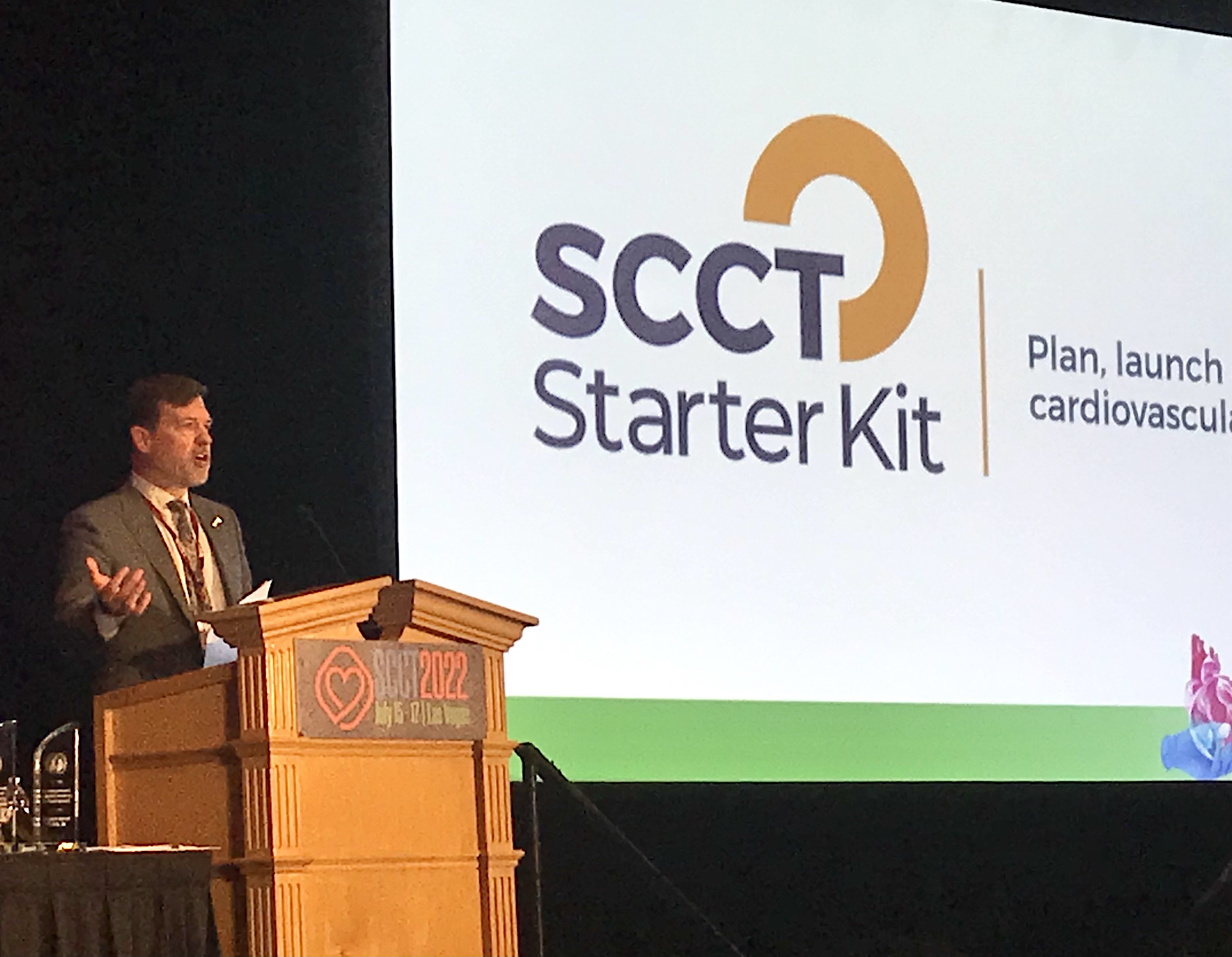
SCCT 2021-22 President Eric Williamson, MD, Mayo Clinic, explains some of the advances with SCCT, including the creation of a new website to help clinics and hospitals start cardiac CT programs. The SCCT Starter Kit site offers information on what hardware, training and accreditation is needed, basic CT scanner specifications, and other resources needed to get started. He said the society saw a rapid uptick in interest in CTA since it was included as a 1A indication for front line imaging in the 2021 Chest Pain Evaluation Guidelines.
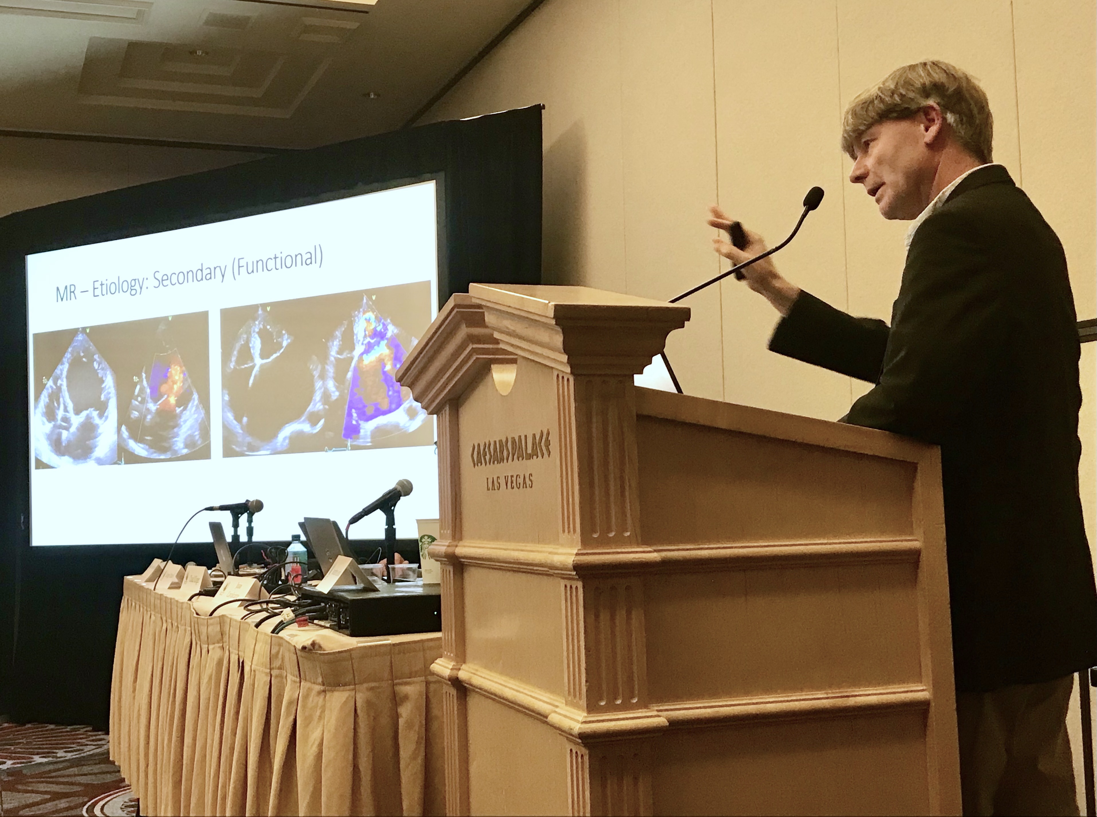
Former European Society of Cardiology (ESC) president Prof. Jeroen Bax, director of noninvasive imaging and director of the echo laboratory, Department of Cardiology at the Leiden University Medical Center in The Netherlands, explains the etiology of secondary mitral regurgitation during a hands-on, how-to course for cardiac CT structural heart assessments and procedural planning. Watch a VIDEO interview with Bax at SCCT 2022.
AI-generated coronary tree from a patient's CT scan showing a color code of areas of interest for plaque burden from the Cleerly software shown at SCCT 2022. This is one of several views created by this FDA-cleared artificial intelligence application in its automated reports. Read more on this trend in the article Cardiac CT soft plaque assessment my offer paradigm shift for coronary disease screening.
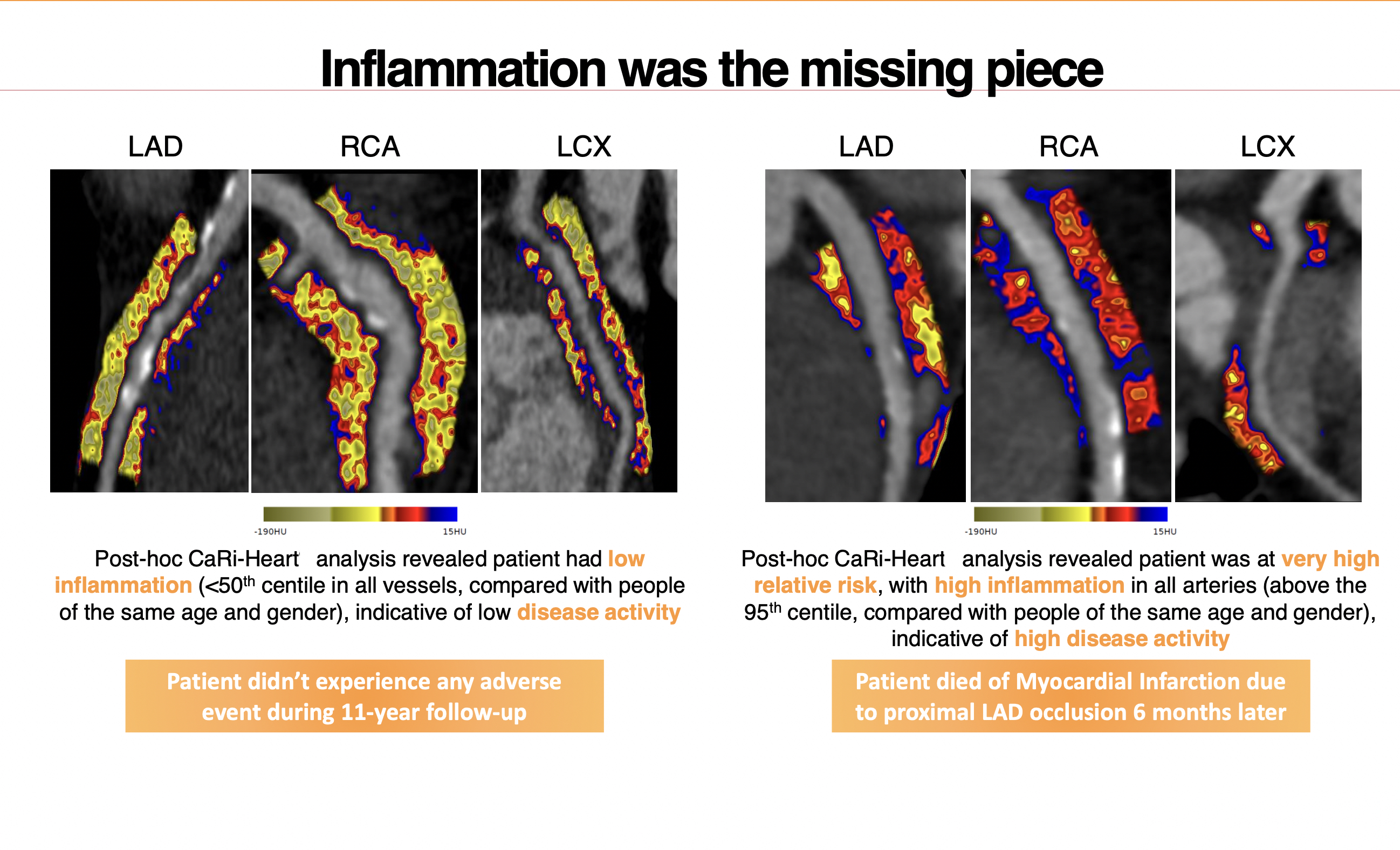
Examples of Caristo's fat attenuation index (FAI) CT imaging of coronary vessel fat to show inflammation. It uses the fat as an indicator if a coronary plaque is inflamed to provide additional information on risk of cardiac events and the progression of vulnerable plaques. It also can show reduction in inflammation due to medical therapy. This can be used to serial scan cardiac patients to better track their disease, better manage drug and lifestyle changes, and enable preventive care years before symptoms appear. The vendor has European approval for the product and is seeking FDA clearance.
One of several pages of an artificial intelligence report on the coronary plaque from a patient's CTA scan. The FDA-cleared software developed Cleerly enables rapid soft plaque AI assessment, overcoming the previously tedious and time consuming manual task of making these calculations. These types of reports may enable a new level of preventive care in cardiology, treating patients long before they have symptomatic disease. Read more on this trend in the article Cardiac CT soft plaque assessment my offer paradigm shift for coronary disease screening.
Kelley Branch, MD, MSc, FSCCT, associate director, clinical trials service unit, professor, of medicine, University of Washington Medical Center, helps an attendee learning how to evaluate cardiac CT congenital heart cases. The Congenital Disease Workshop as an all-day pre-conference course to sharpen reading skills for these difficult to interpret cases.
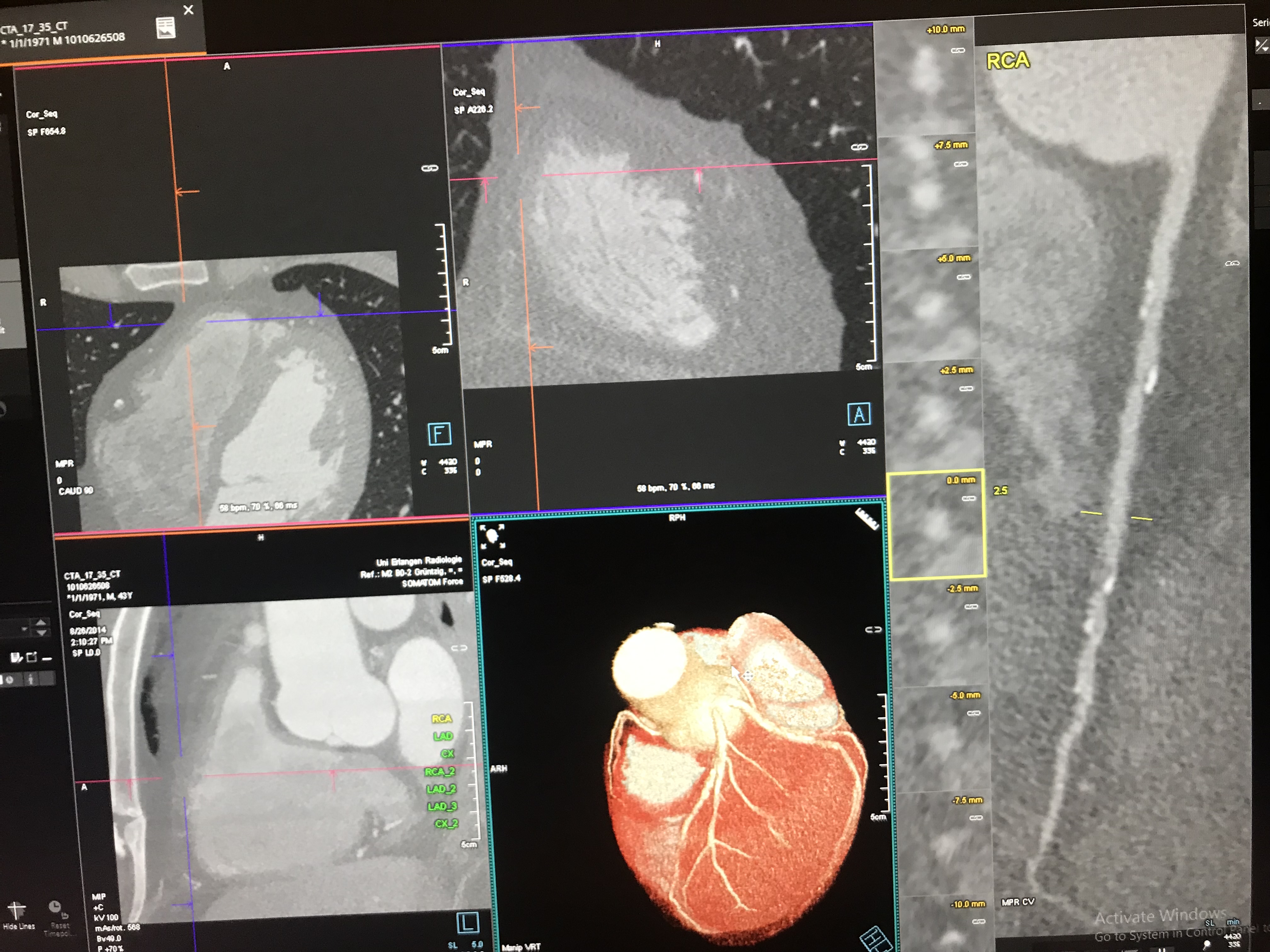
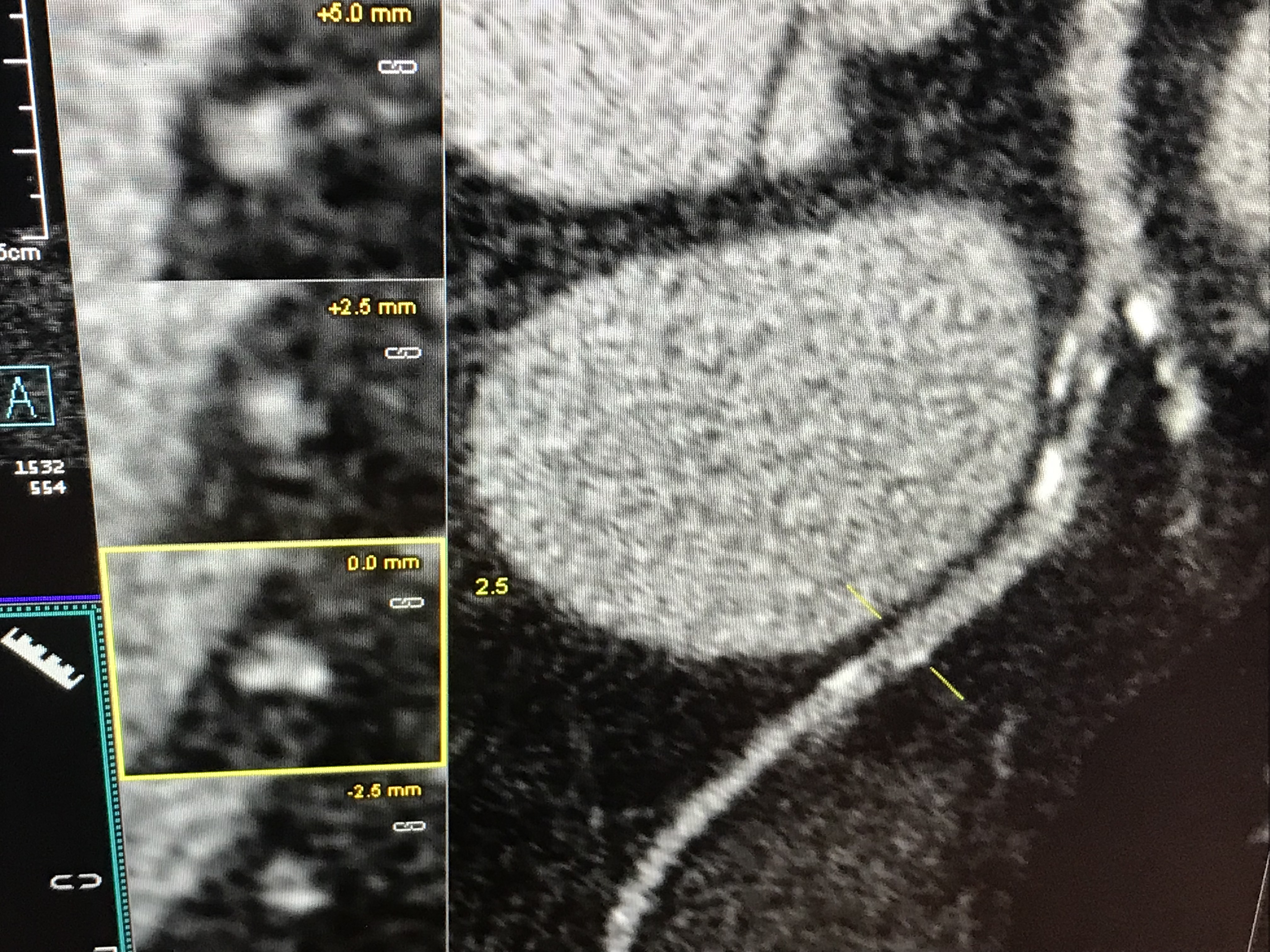
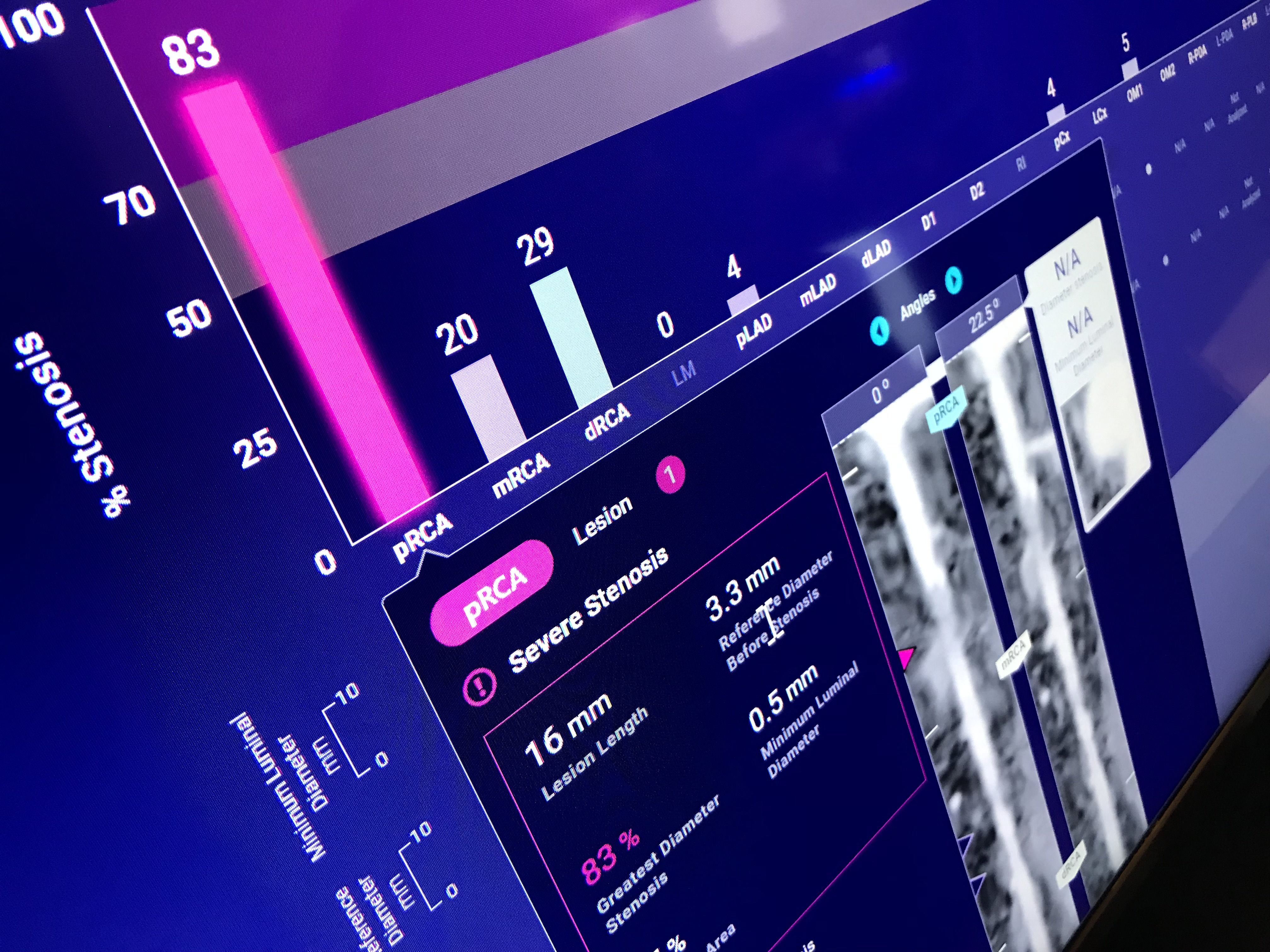
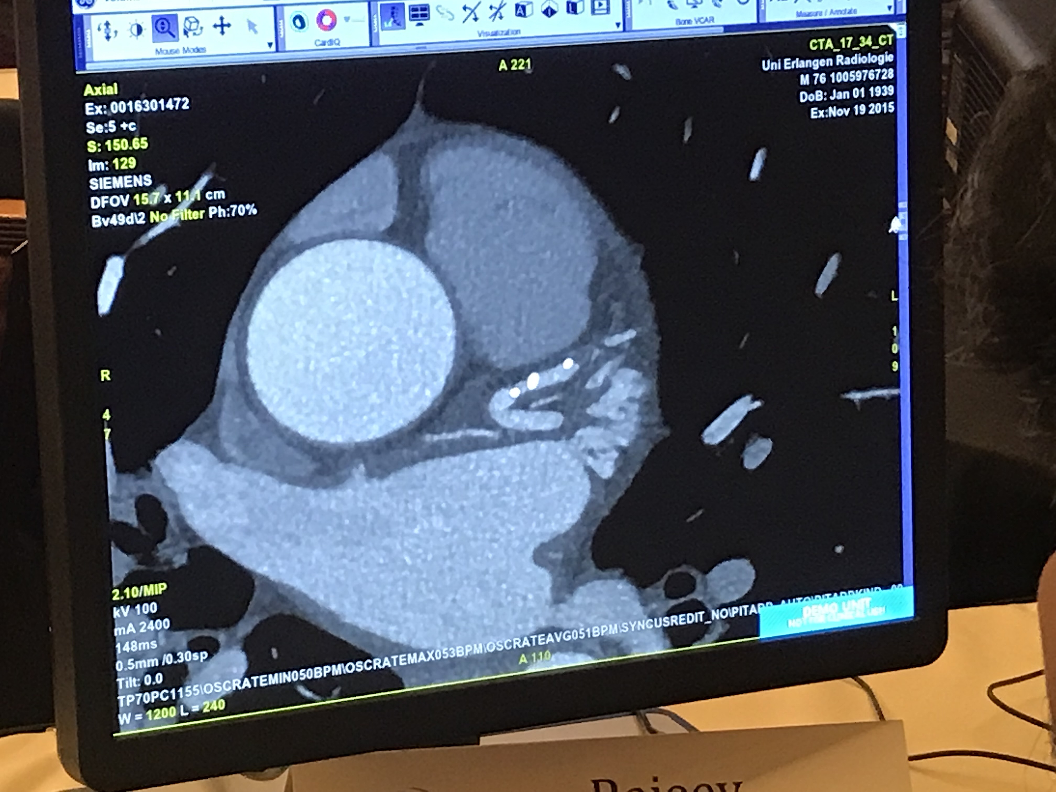
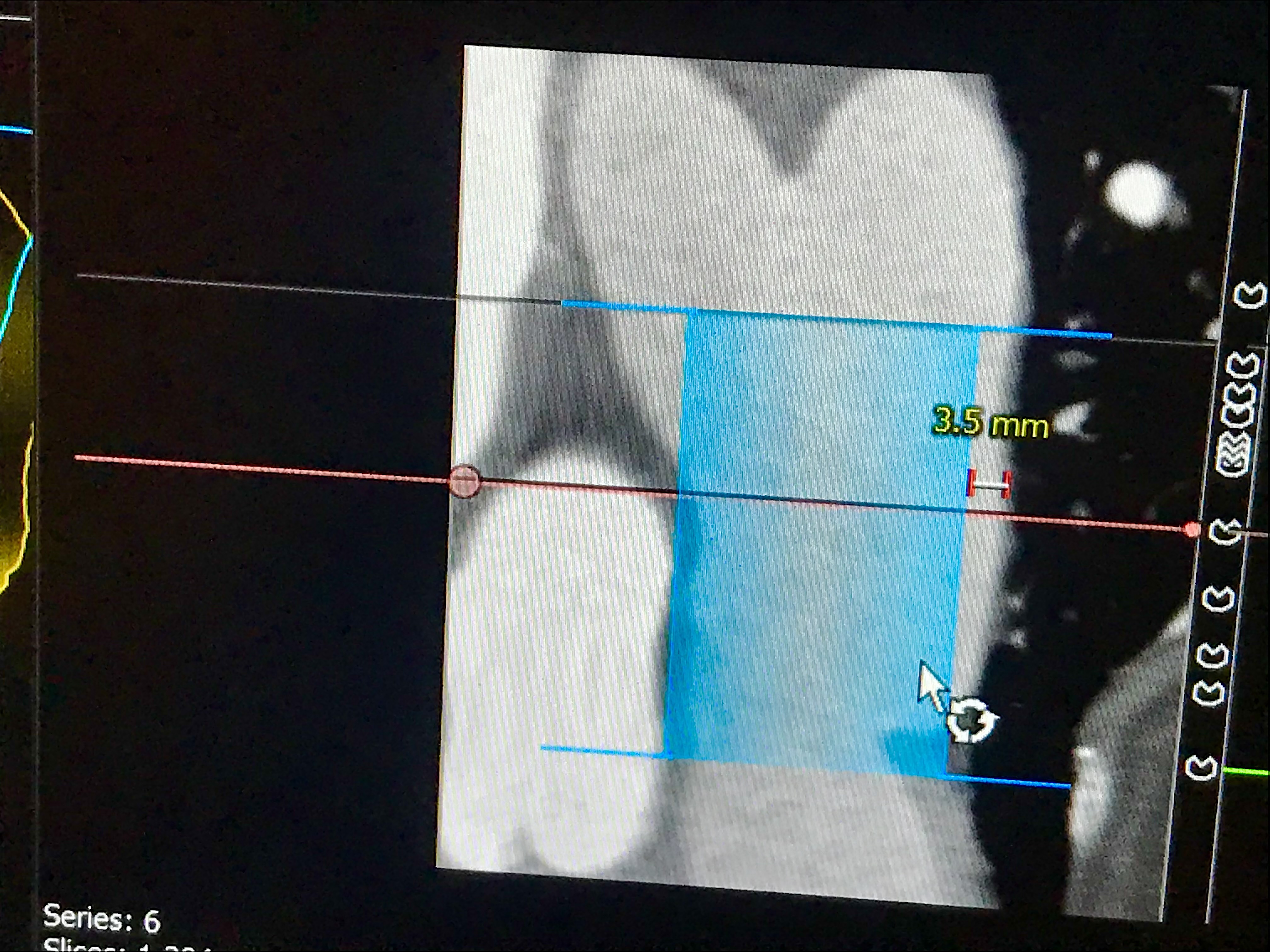
Coronary CT angiography (CCTA) exam showing the heart in three different planes, a 3D reconstruction and a elongated reconstructed view of a coronary artery. The stretched artery view allows assessment on plaque inside the vessel and the small thumbnail images show cross sectional views of the vessel to better understand the plaque and accompanying vessel remodeling. Example is from a Siemens floor demonstration.
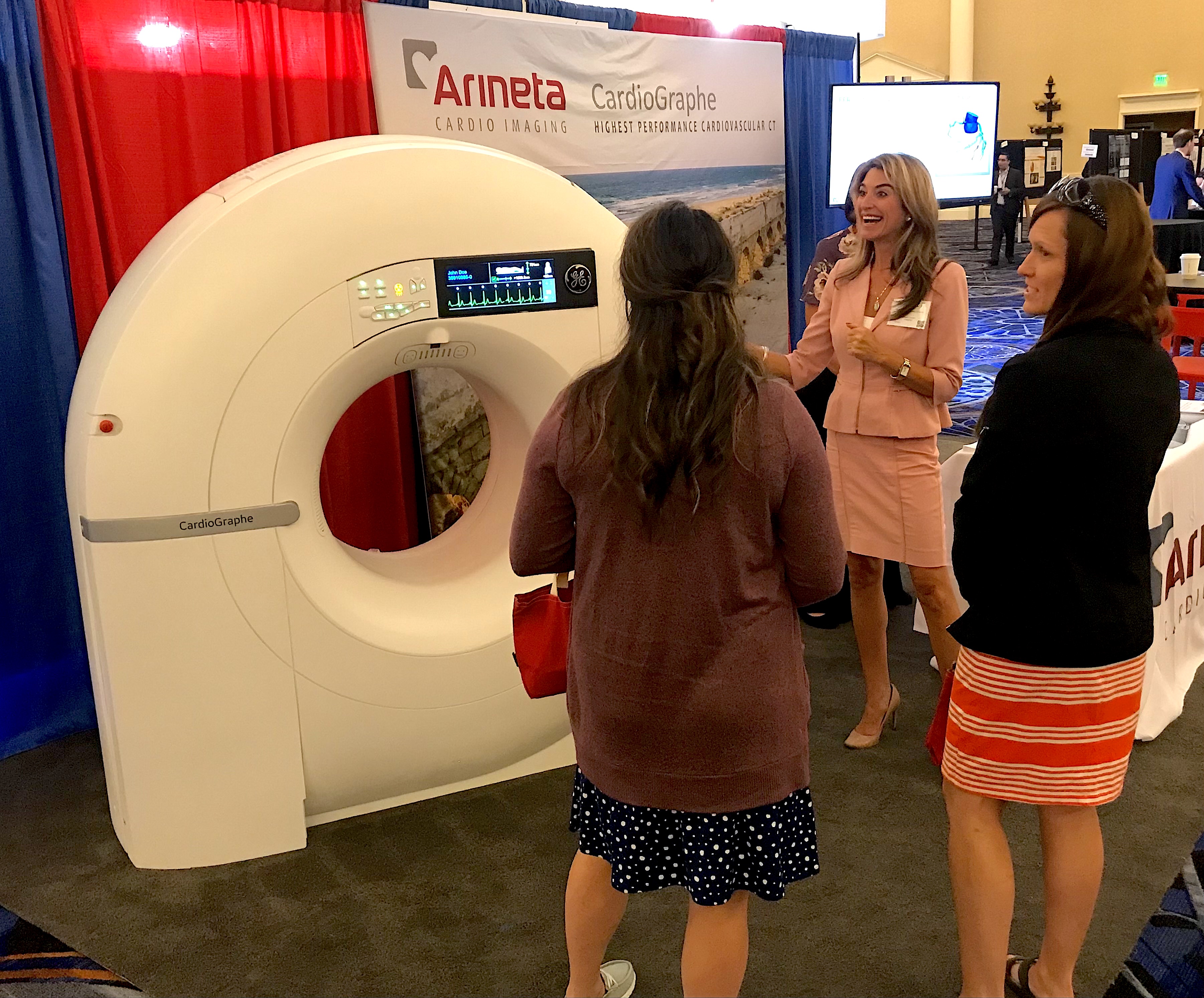
The small Cardiograph dedicated cardiac CT system on display at SCCT is as designed for use in cardiology departments or out patient cardiac clinics. It comes in a price-point lower than conventional CT scanners that are cardiac capible. One of the first outpatient clinics in the U.S. to install this system was Duly Healthcare in the Chicago suburbs. They use it to evaluate chest pain, calcium scoring exams and imaging workups structural heart procedures. Watch the VIDEO: Office-based cardiac CT and FFR-CT offer a new business model, or in this PHOTO GALLERY: Duly Health adopts outpatient cardiac CT as a standard of care.
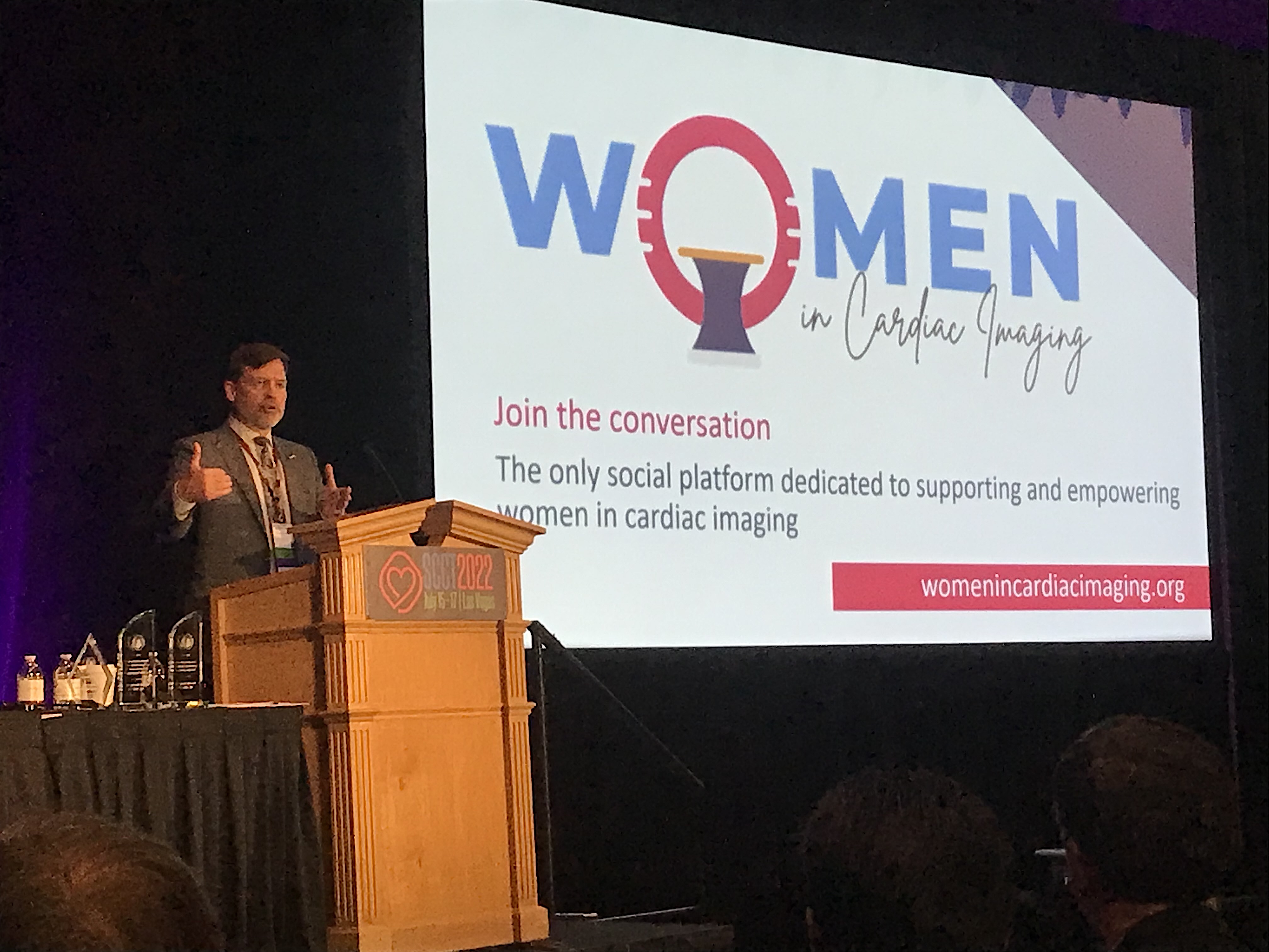
SCCT 2021-22 President Eric Williamson, MD, Mayo Clinic, explains some of the advances with SCCT, including creation of the social media platform "Women in Cardiac Imaging," which is dedicated to supporting and empowering women in the cardiac imaging space.
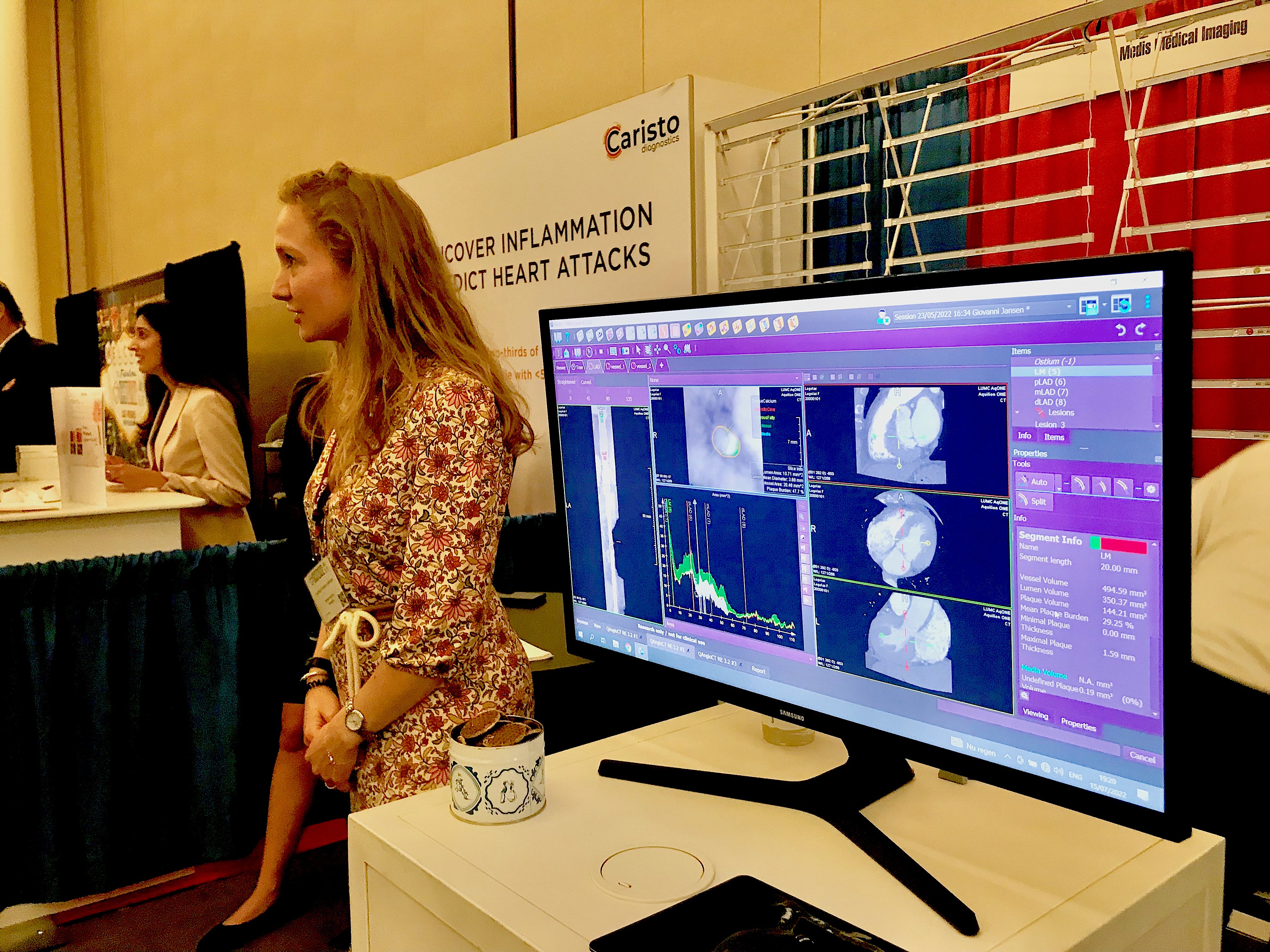
Automated coronary plaque assessment software displayed by Medis on the SCCT expo floor. Read more on this trend in the article Cardiac CT soft plaque assessment my offer paradigm shift for coronary disease screening.
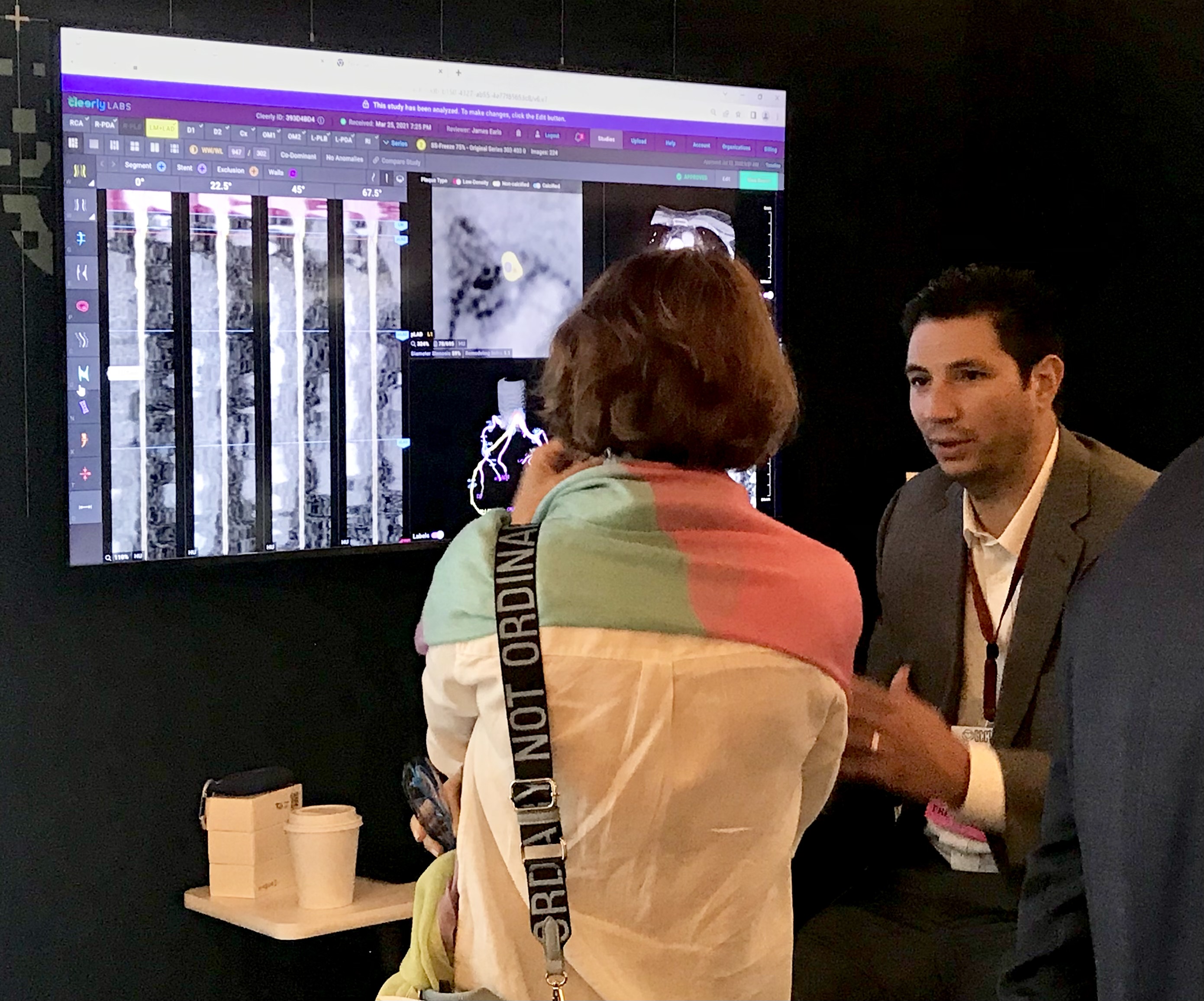
Discussion on the expo floor on how artificial intelligence can help assess coronary soft plaque in the Cleerly booth. The vendor has FDA clearance for its software. Read more in the article Cardiac CT soft plaque assessment my offer paradigm shift for coronary disease screening.
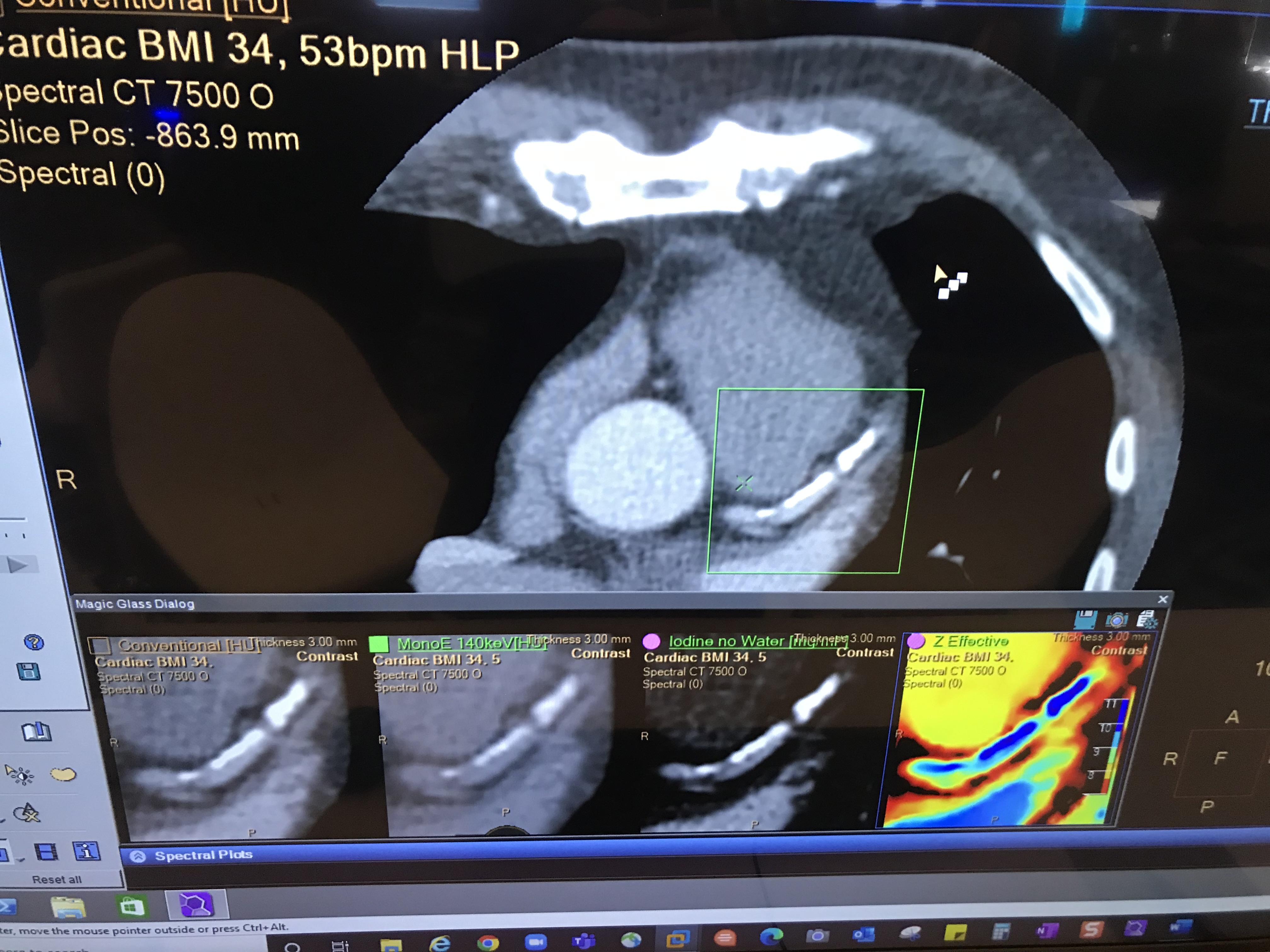
Example of CT spectral imaging from Philips Healthcare, showing a heavily calcified coronary vessel at different energies so the reader can see which offers the best view past the calcium.
Pre-conference all-day training course for the Coronary CTA Academy: Cases from Basic to Intermediate. CTA experts explained step by step how to review cardiac CT scans and how to do basic post-processing of the datasets.
Example of a coronary artery CT angiography (CTA) assessment using curved MPR image to show the entire length of the vessel in one image. Example shown by Siemens.
One of the packed sessions at SCCT on new photon-counting CT technology.
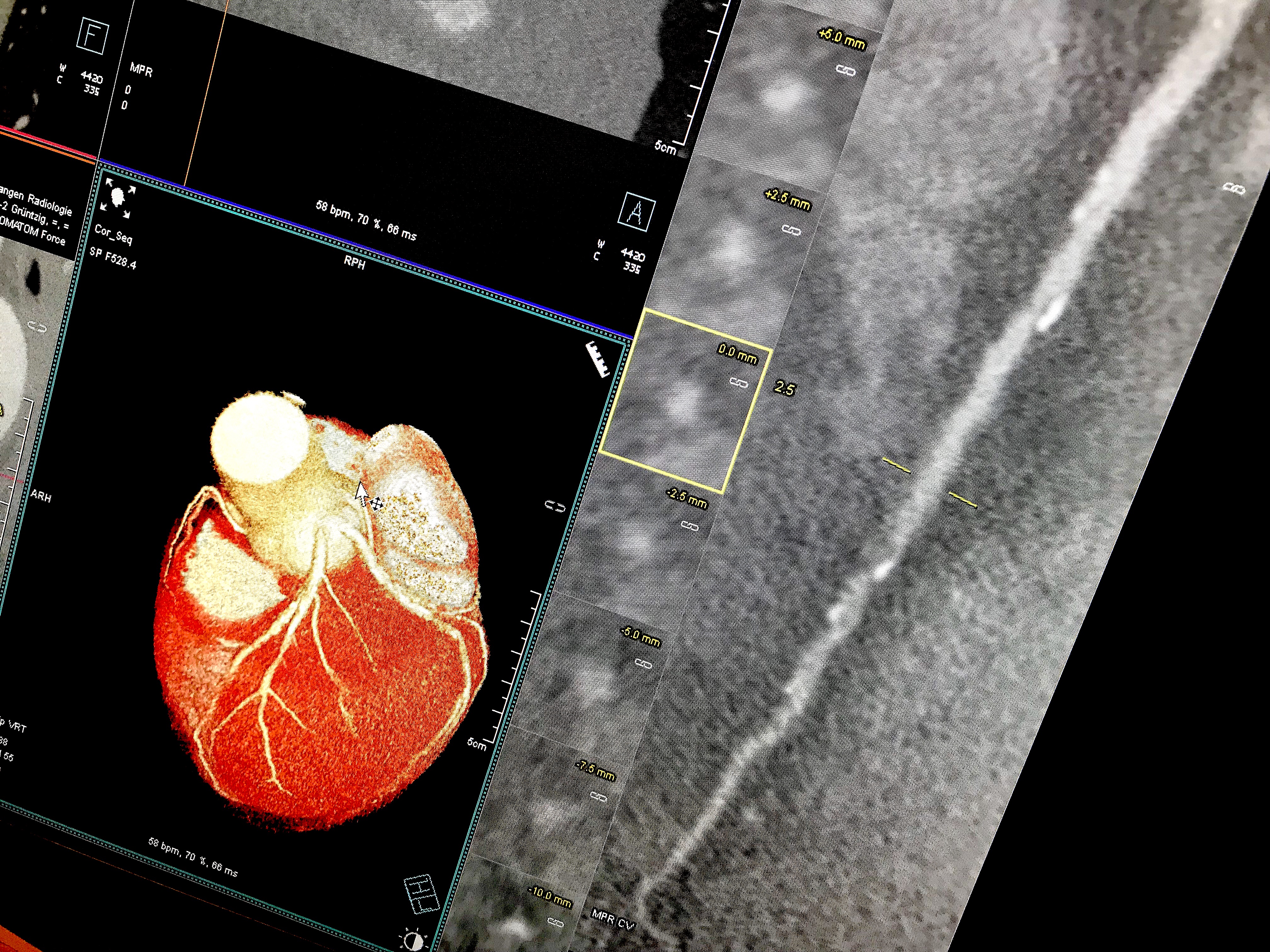
Example of a curved MPR image reconstruction of entire length of a coronary artery on a cardiac CT scan to better show calcified and soft plaque burden inside the vessel. The thumbnail dots on the left side of the image are cross sectional views of the vessel. Siemens example on the expo floor.
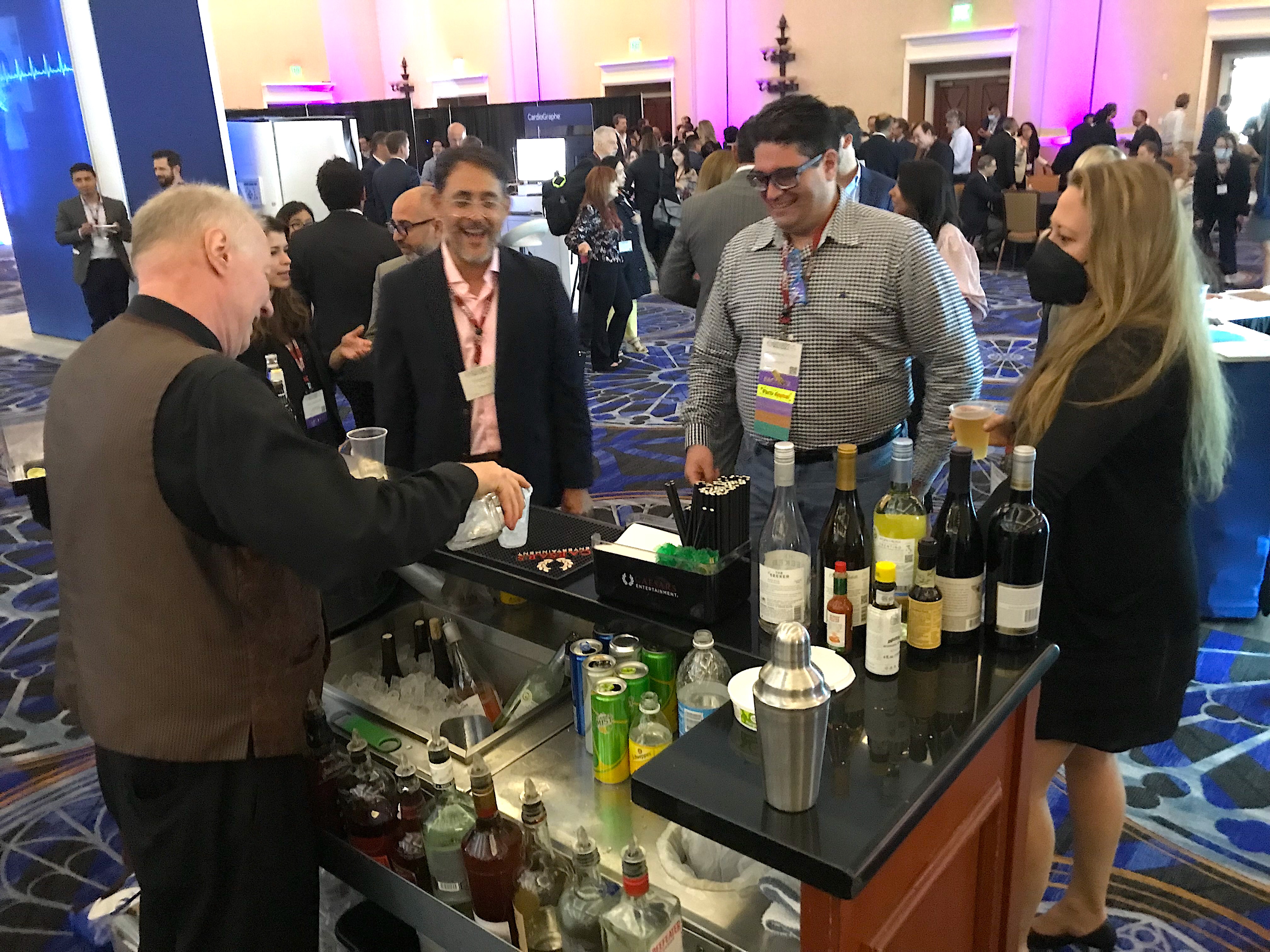
Attendees getting drinks on the expo floor during an evening reception during SCCT. Many attendees said they were glad to be back in person for networking, after two years of COVID required virtual meetings in 2020 and 2021.

A SCCT attendee taking a break between sessions just outside the meeting rooms overlooking the vast Caeser's Palace pool.
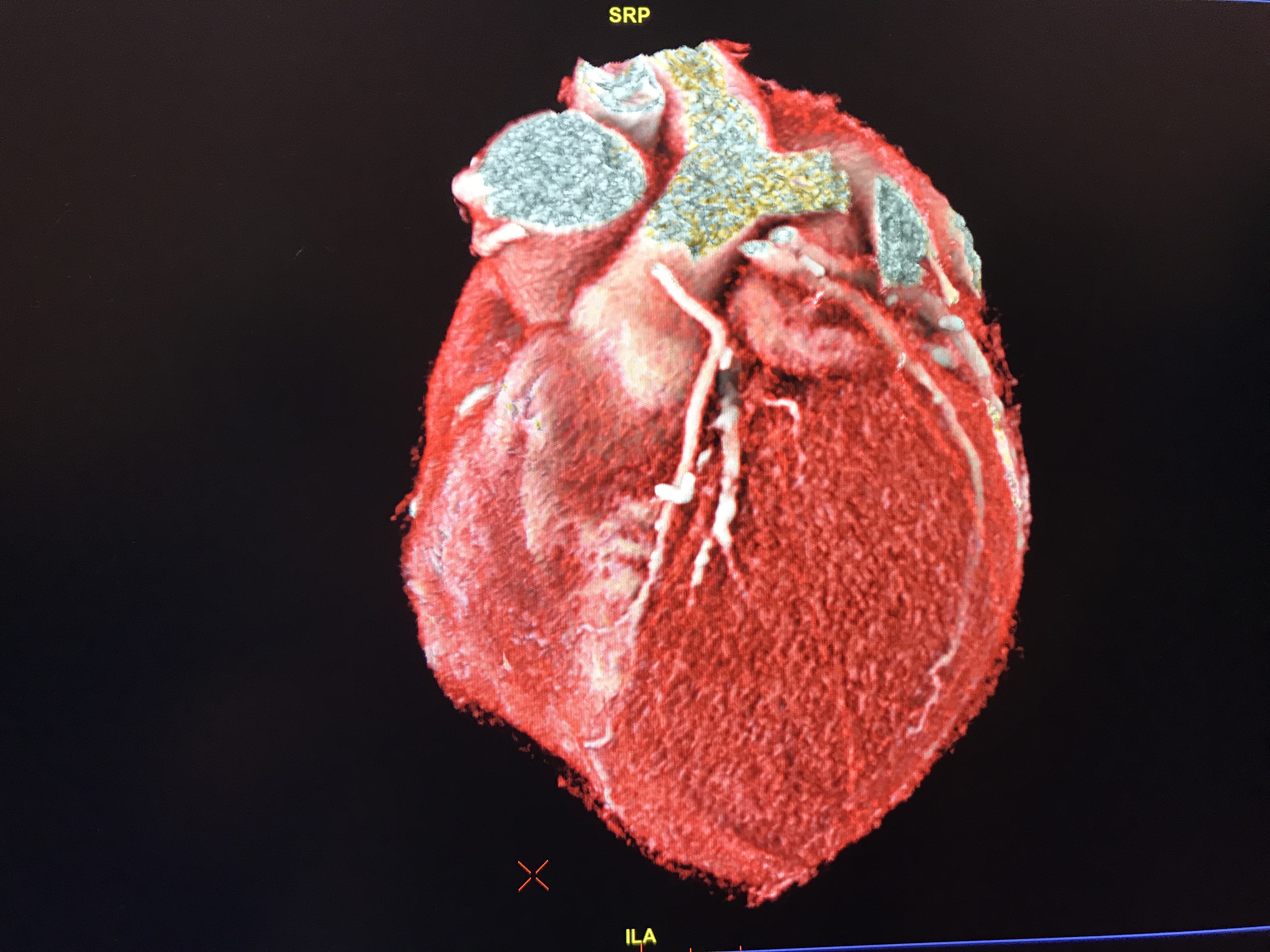
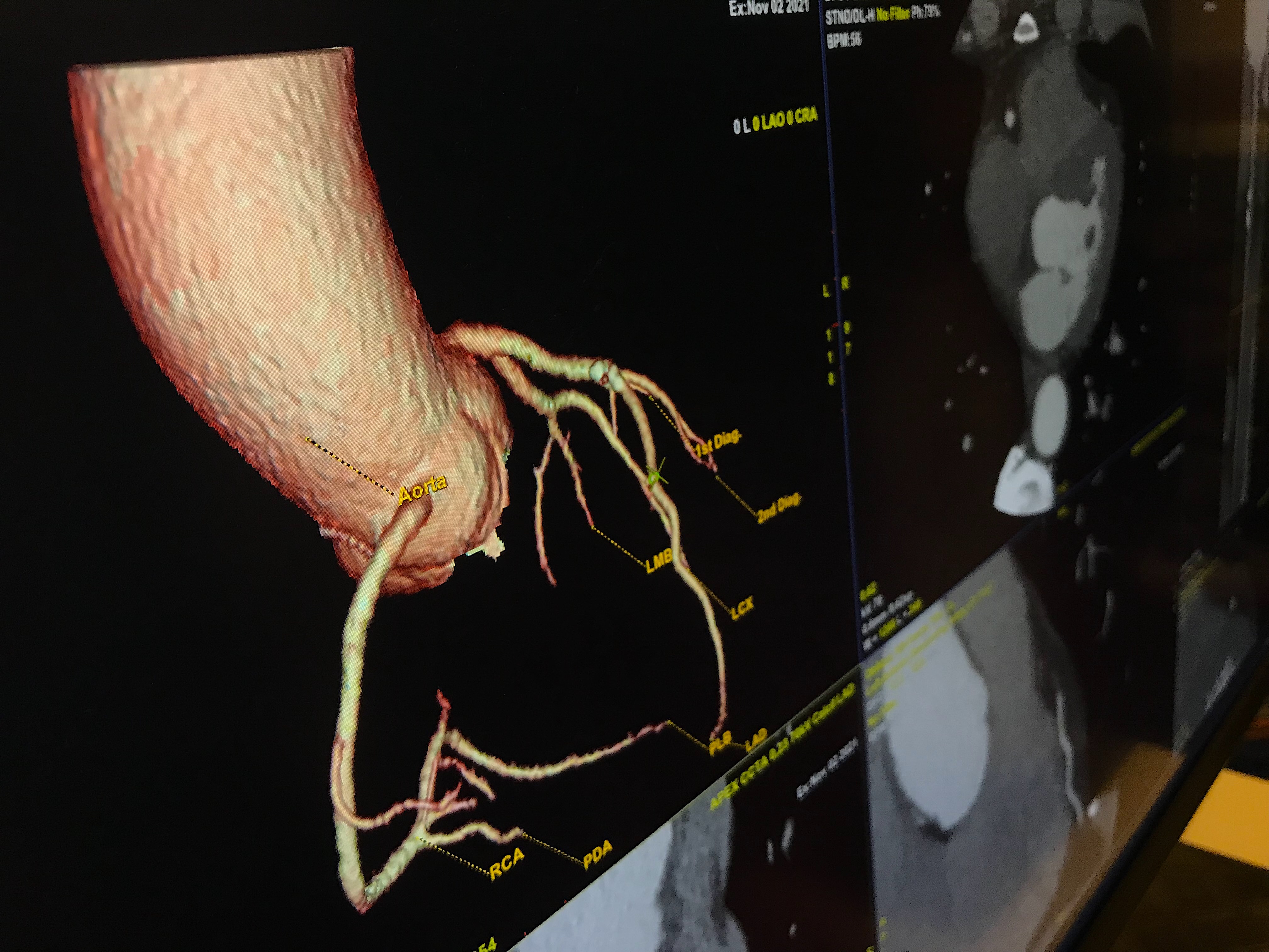
A 3D rendering from a cardiac CT of a patient with a coronary artery bypass graft (CABG). Part of the imaging shown by GE Healthcare from its Revolution Apex CT scanner on the expo floor.
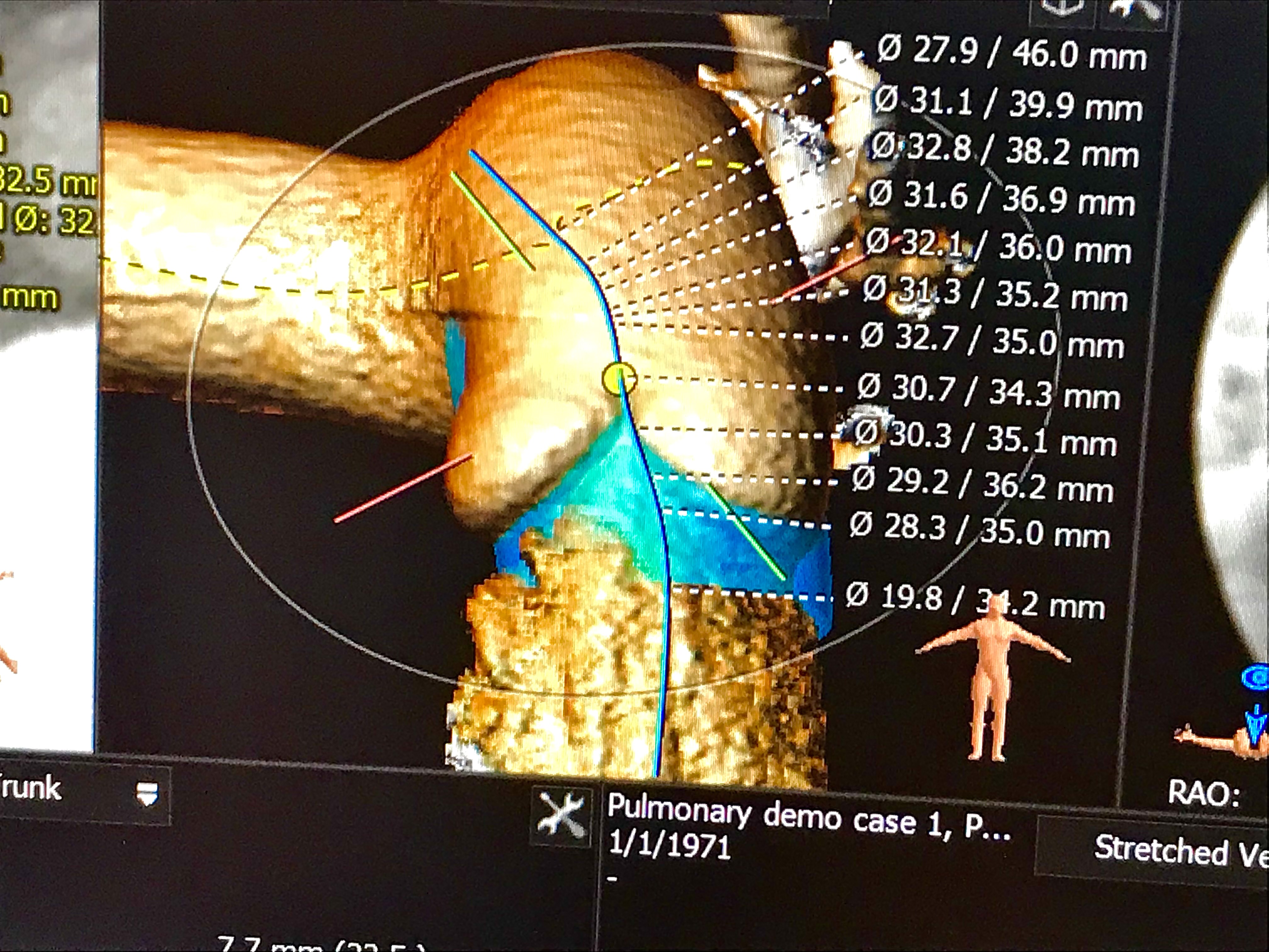
Virtual implantation of a pulmonary valve on CT pre-procedural assessment software in a session on structural heart planning at the Society of Cardiovascular Computed Tomography (SCCT) 2022 meeting in Las Vegas. The software used in this session was 3mensio, which enables the imager to find the center lines of the vessels and valves, measure the annulus and to perform a virtual implant of various commercial valves (Melody, Harmony, Sapien) of different sizes to see how the valve fits in the anatomy. This can reduce procedure time and help in the prior valve sizing.
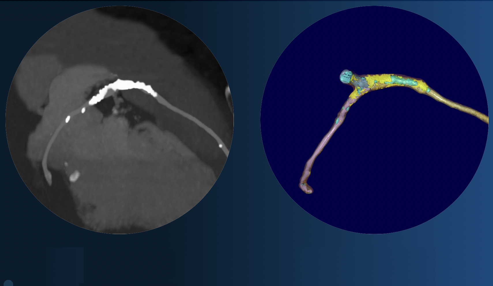
An unusual finding on a cardiac CTA showing the radio-opaque markers for the ends of an Absorb bioresorbable stent, where the stent has completely dissolved, leaving behind only the markers. In the treated area is a calcified lesion showing where the ruptured plaque healed. The stent is no longer on the market due to low usage.
Pre-conference all-day how-to training course for the Coronary CTA Academy: Cases from Basic to Intermediate. CTA experts explained step by step how to review cardiac CT scans and how to do basic post-processing of the datasets.
A SCCT 2022 meeting pre-conference, all-day training course for the Coronary CTA Academy: Cases from Basic to Intermediate. CTA experts explained step-by-step how to review cardiac CT scans and how to do basic post-processing of the datasets.
Kelley Branch, MD, MSc, FSCCT, associate director, clinical trials service unit, professor, of medicine, University of Washington Medical Center, helps an attendee learning how to evaluate cardiac CT congenital heart cases. The Congenital Disease Workshop as an all-day pre-conference course to sharpen reading skills for these difficult to interpret cases.
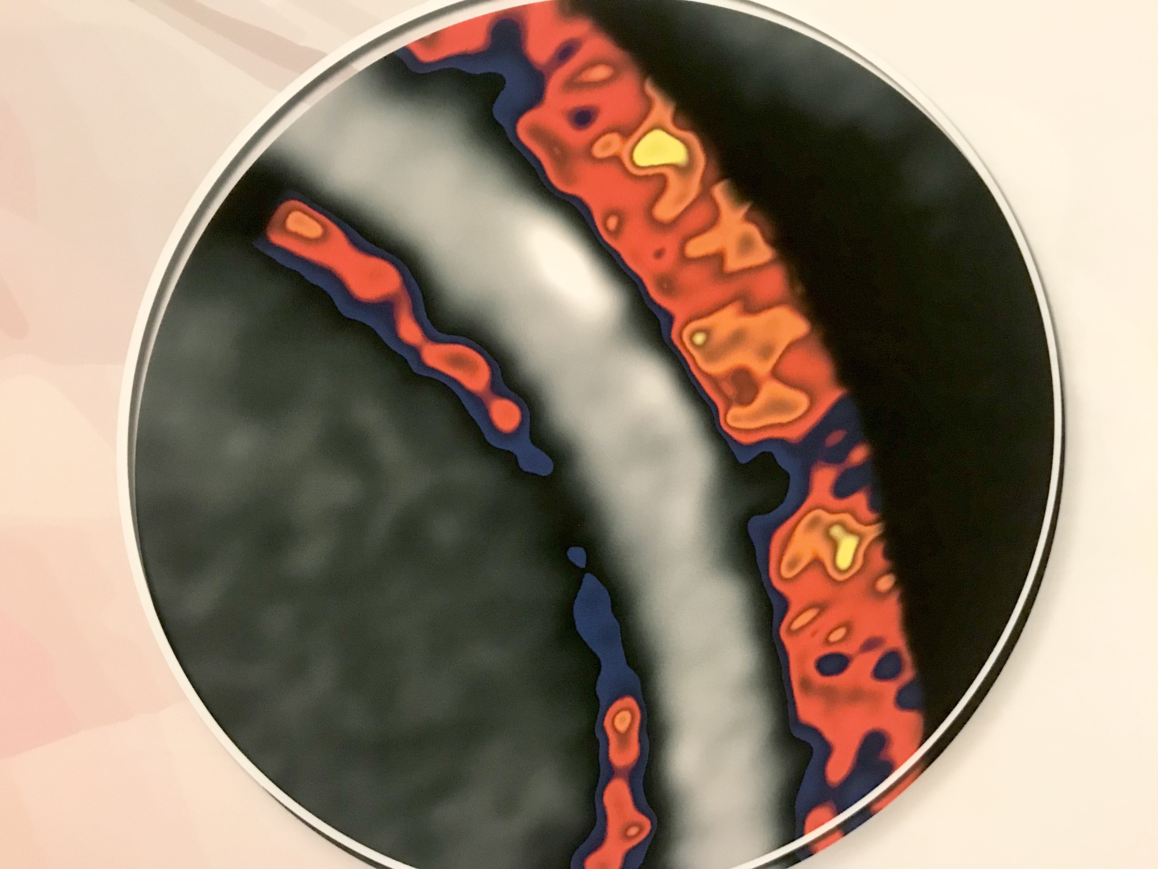
An example of heavy coronary artery inflammation (shown in red) using Caristo's fat attenuation index (FAI) CT imaging assessment software. It shows inflammation in the fat surrounding the coronary vessels as an indicator if a coronary plaque is inflamed to provide additional information on risk of cardiac events and the progression of vulnerable plaques. The technology has CE mark and they are seeking FDA.
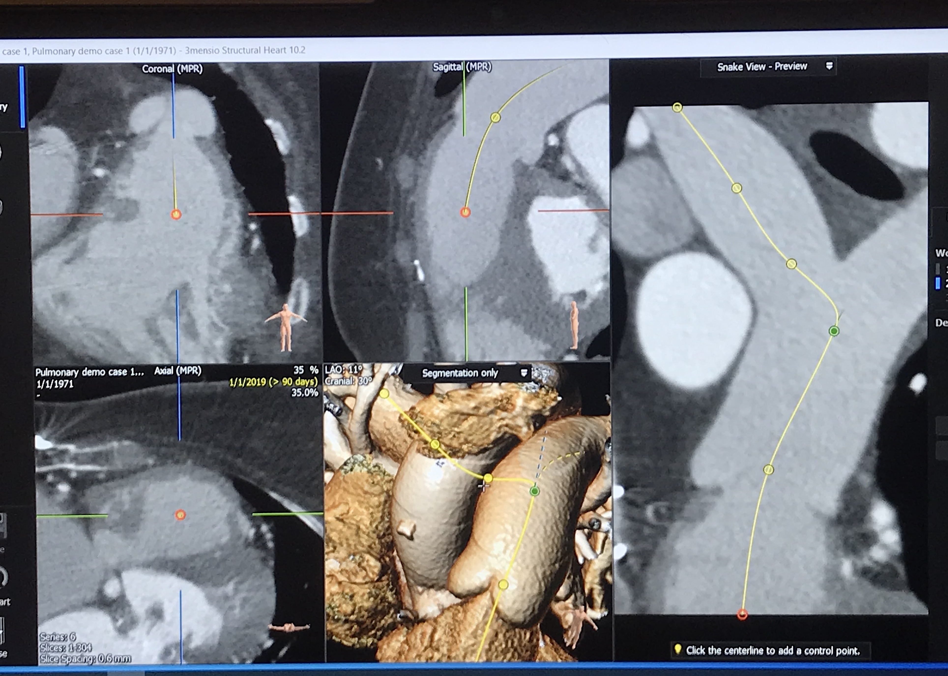
CT planning case for implantation of a transcatheter a pulmonary valve on CT pre-procedural assessment in a session on structural heart planning at the Society of Cardiovascular Computed Tomography (SCCT) 2022 meeting last week. The software used in this session was 3mensio, which enables the imager to find the center lines of the vessels and valves, measure the annulus and to perform a virtual implant of various commercial valves (Melody, Harmony, Sapien) of different sizes to see how the valve fits in the anatomy. This can reduce procedure time and help in the prior valve sizing.
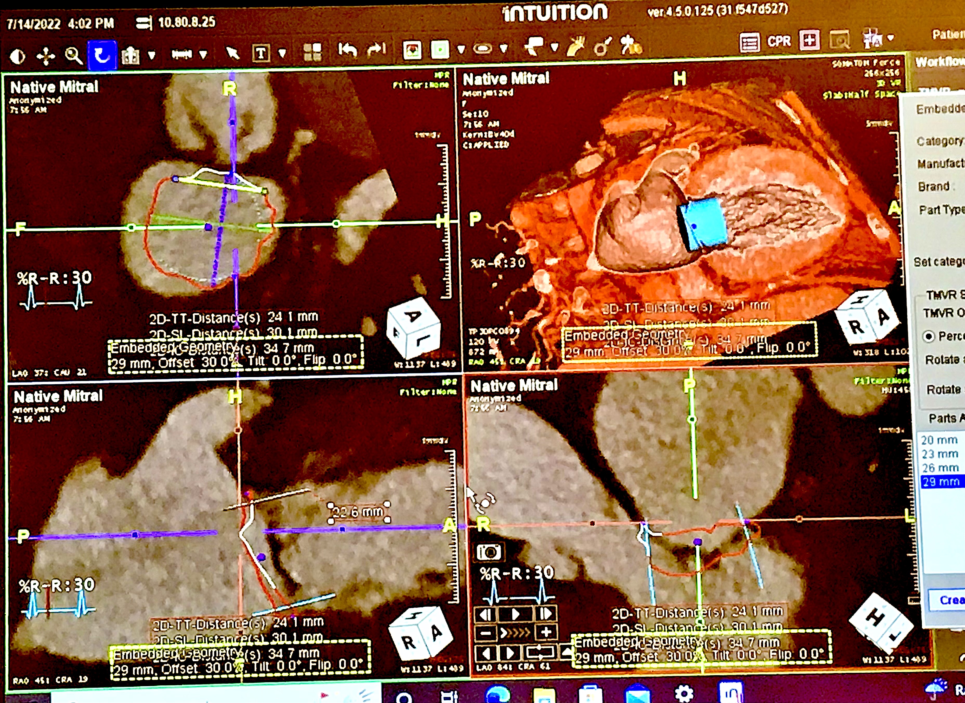
Virtual implantation of a mitral valve using the Intuition advanced visualization software during CT pre-procedural structural heart planning seminar at the Society of Cardiovascular Computed Tomography (SCCT) 2022 meeting. Use of virtual implants is growing to get a better idea of how different size devices fit the anatomy and what complications they might present, such as left ventricular outflow tract (LVOT) obstruction, and to calculate neo-LVOT area in mitral valve procedures.
Virtual implantation of a pulmonary valve on CT pre-procedural assessment software in a session on structural heart planning at the Society of Cardiovascular Computed Tomography (SCCT) 2022 meeting in Las Vegas. The software used in this session was 3mensio, which enables the imager to find the center lines of the vessels and valves, measure the annulus and to perform a virtual implant of various commercial valves (Melody, Harmony, Sapien) of different sizes to see how the valve fits in the anatomy. This can reduce procedure time and help in the prior valve sizing.
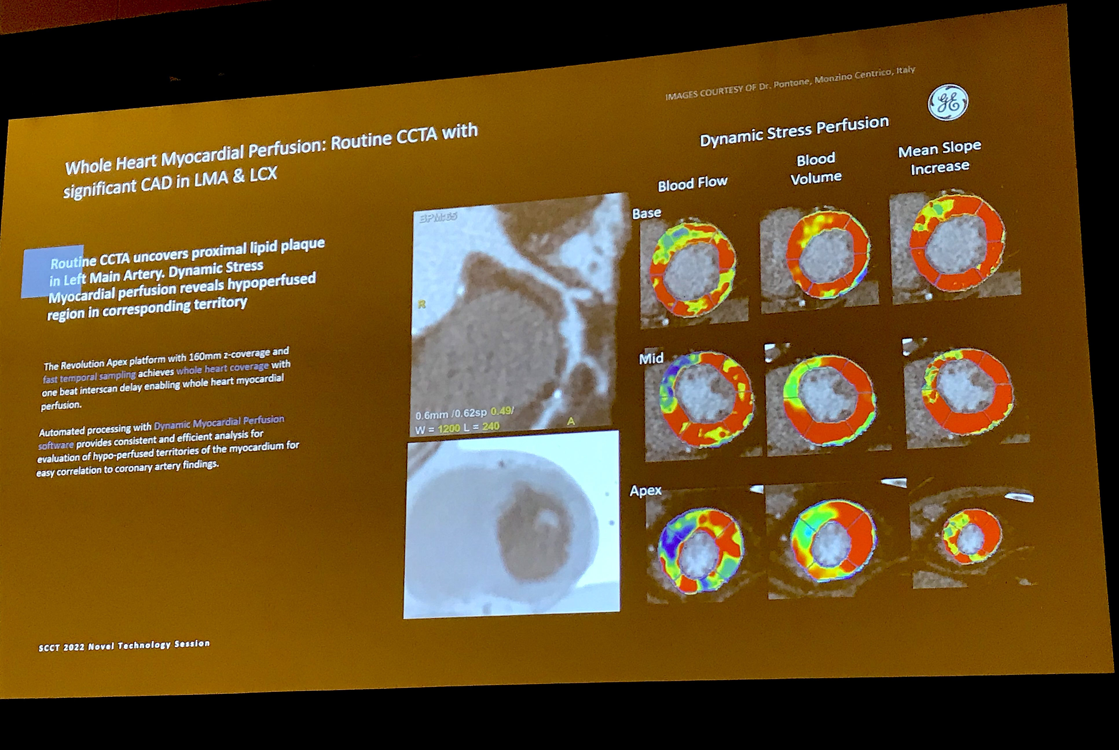
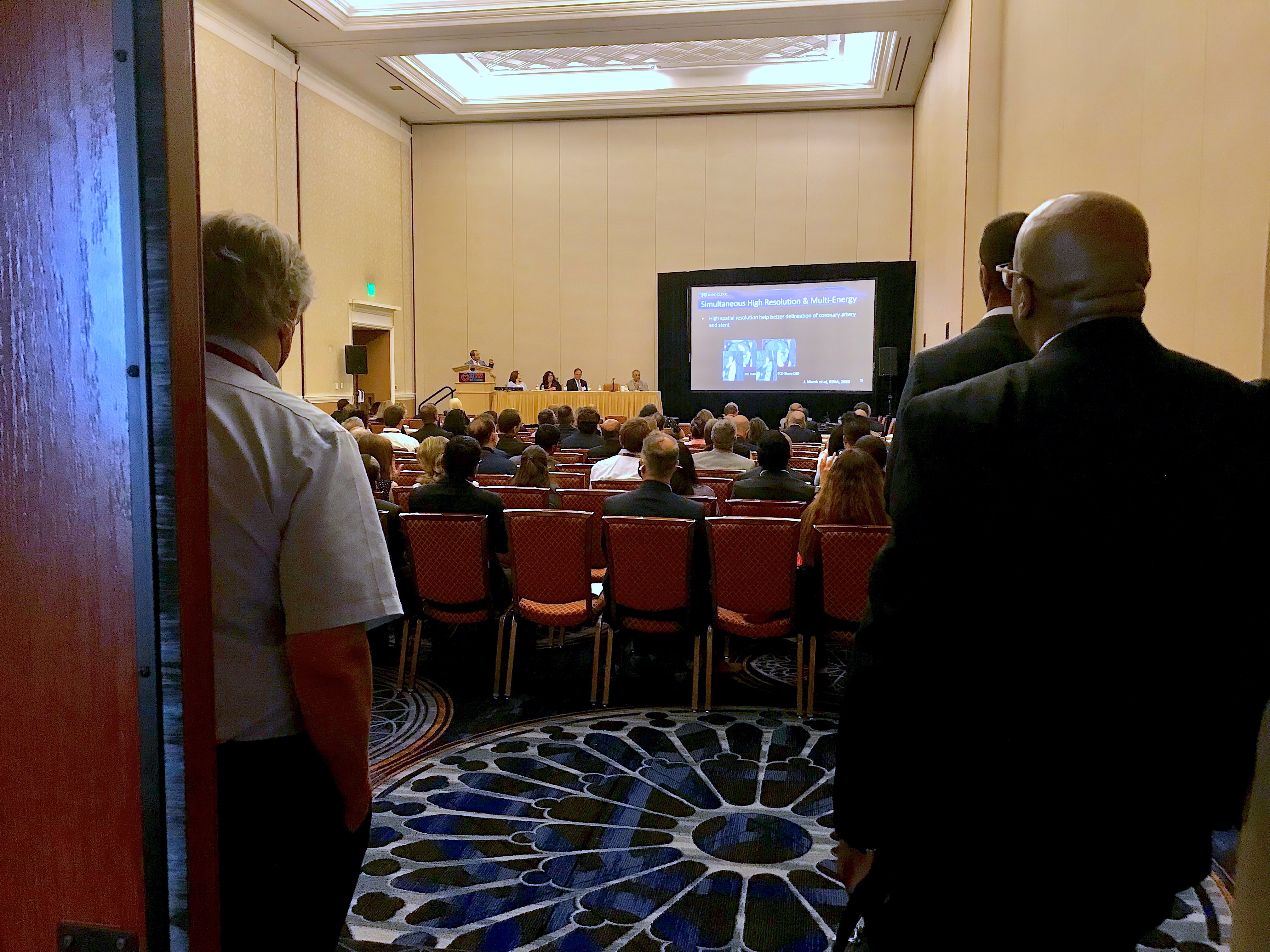
Presentation slide from a new cardiac CT innovations session showing GE Healthcare's whole heart dynamic stress perfusion software the vendor is developing. Vendors are attempting to add more physiological information to CTA so additional tests to examine cardiac function are not needed.
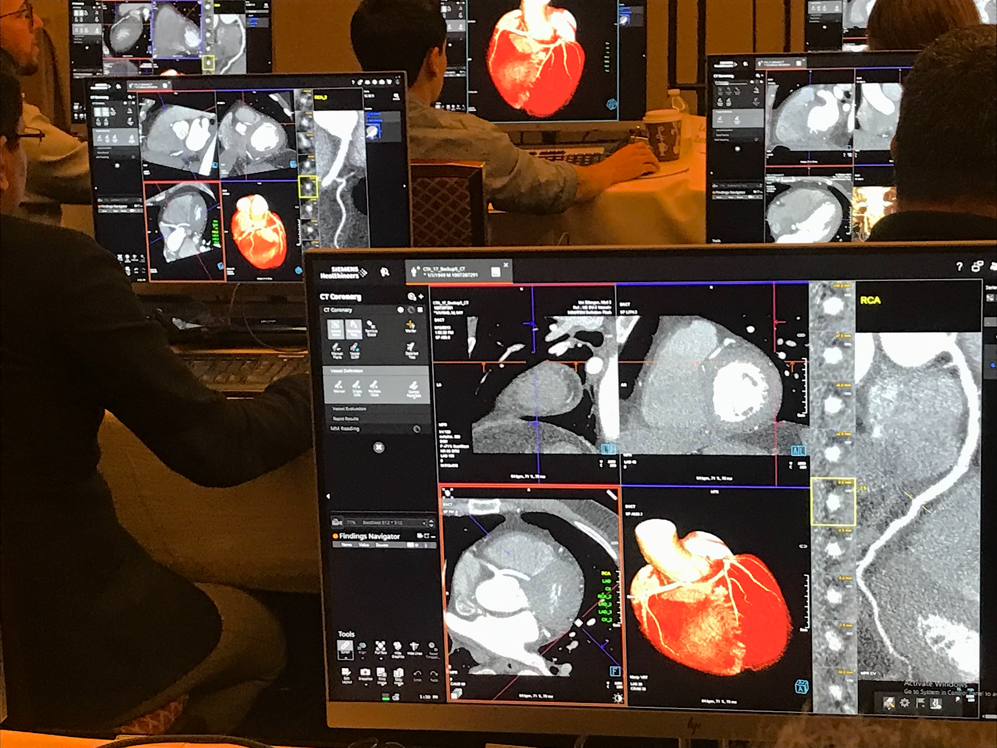
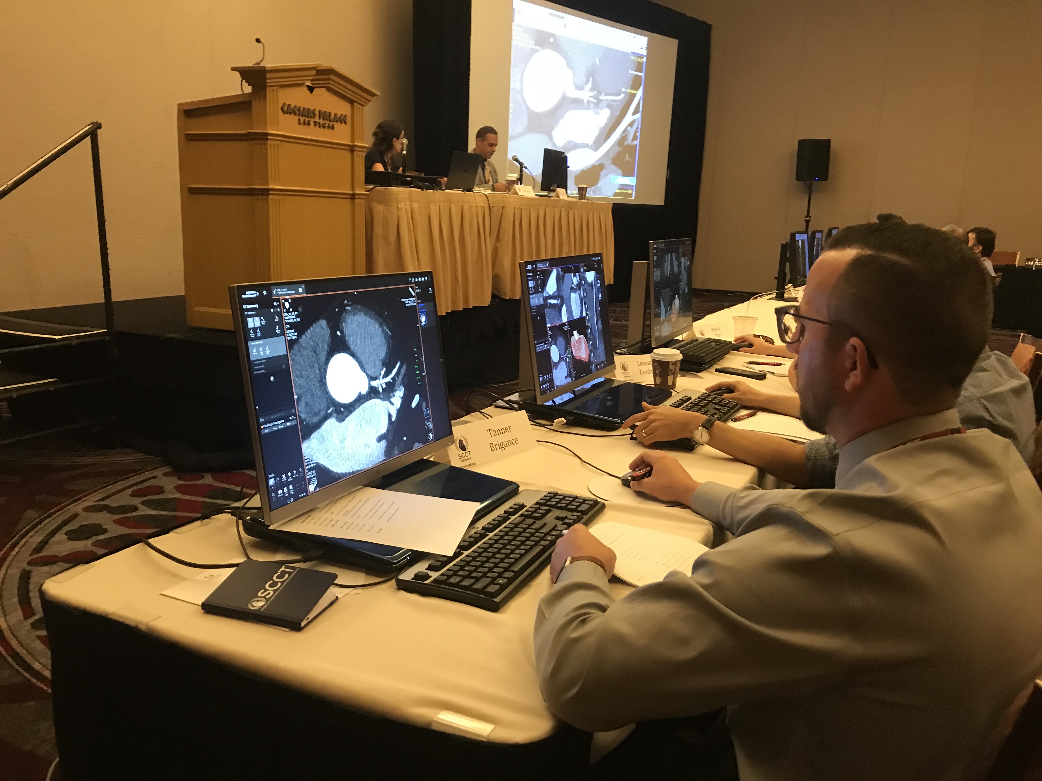
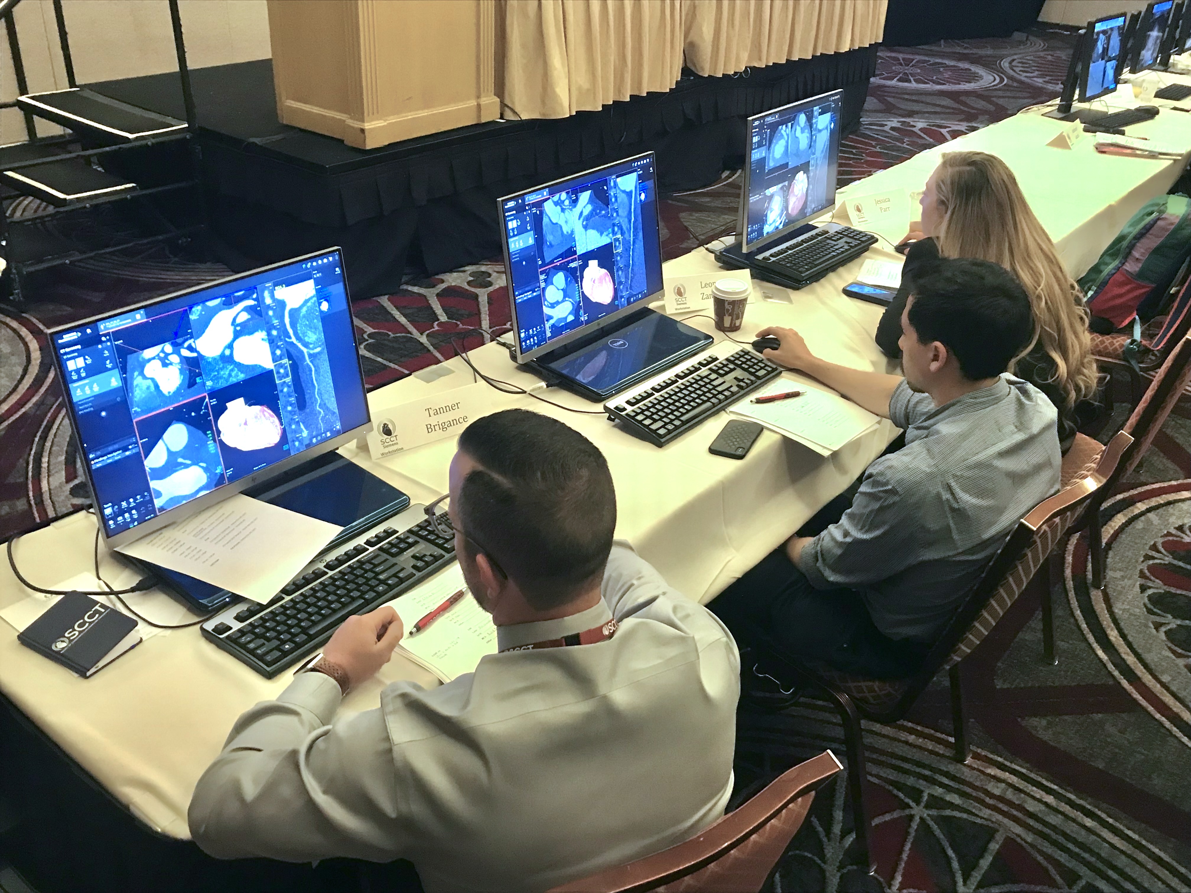
A sea of workstation screens showing cardiac CTA post-processing assessments during a hands-on training session at SCCT.
SCCT President Eric Williams, MD, Mayo Clinic, speaks at the opening session of the SCCT meeting. He explained cardiac CT is doing very well and saw a major boost in interest from the 2021 Chest Pain Guidelines, which elevated cardiac CT to a 1A front line imaging recommendation. Here more about on his overview in the VIDEO: Top 6 takeaways from the Society of Cardiovascular CT 2022 meeting.
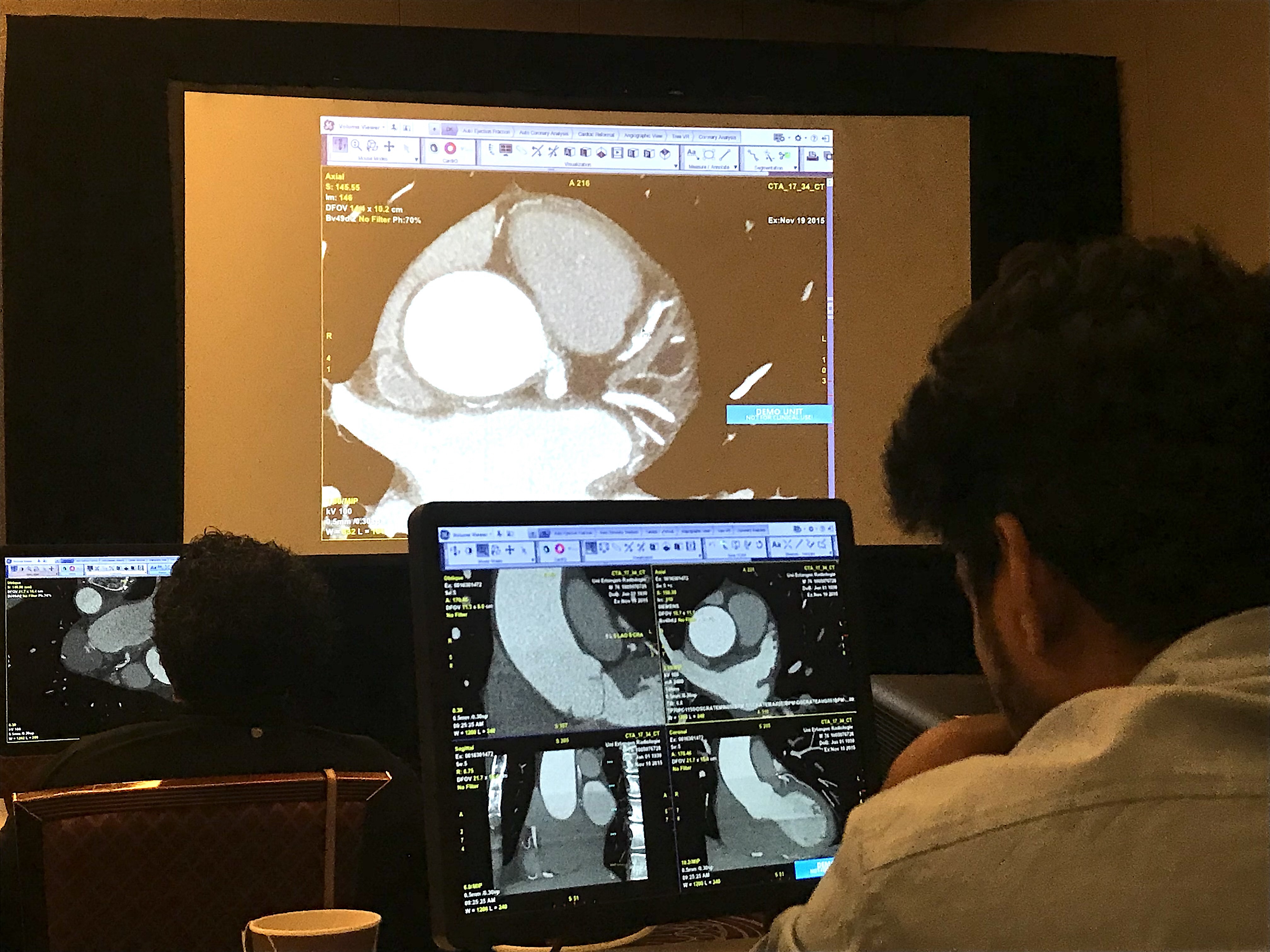
Post-processing of a cardiac CT dataset to extract a 3D view of a patient's coronary arteries. Part of a GE booth demo on the show floor.
Mirvat Alasnag, MD, director of catheterization laboratory, King Fahd Armed Forces Hospital, Jeddah, Saudi Arabia, Board Of Trustees for the Society for Cardiovascular Angiography and Interventions (SCAI), and first female interventional cardiologist in the Gulf region, presents on what the ISCHEMIA trial means for coronary artery disease imaging.
Advanced visualization for CT of the myocardium demonstrated in the Ziosoft booth on the expo floor.

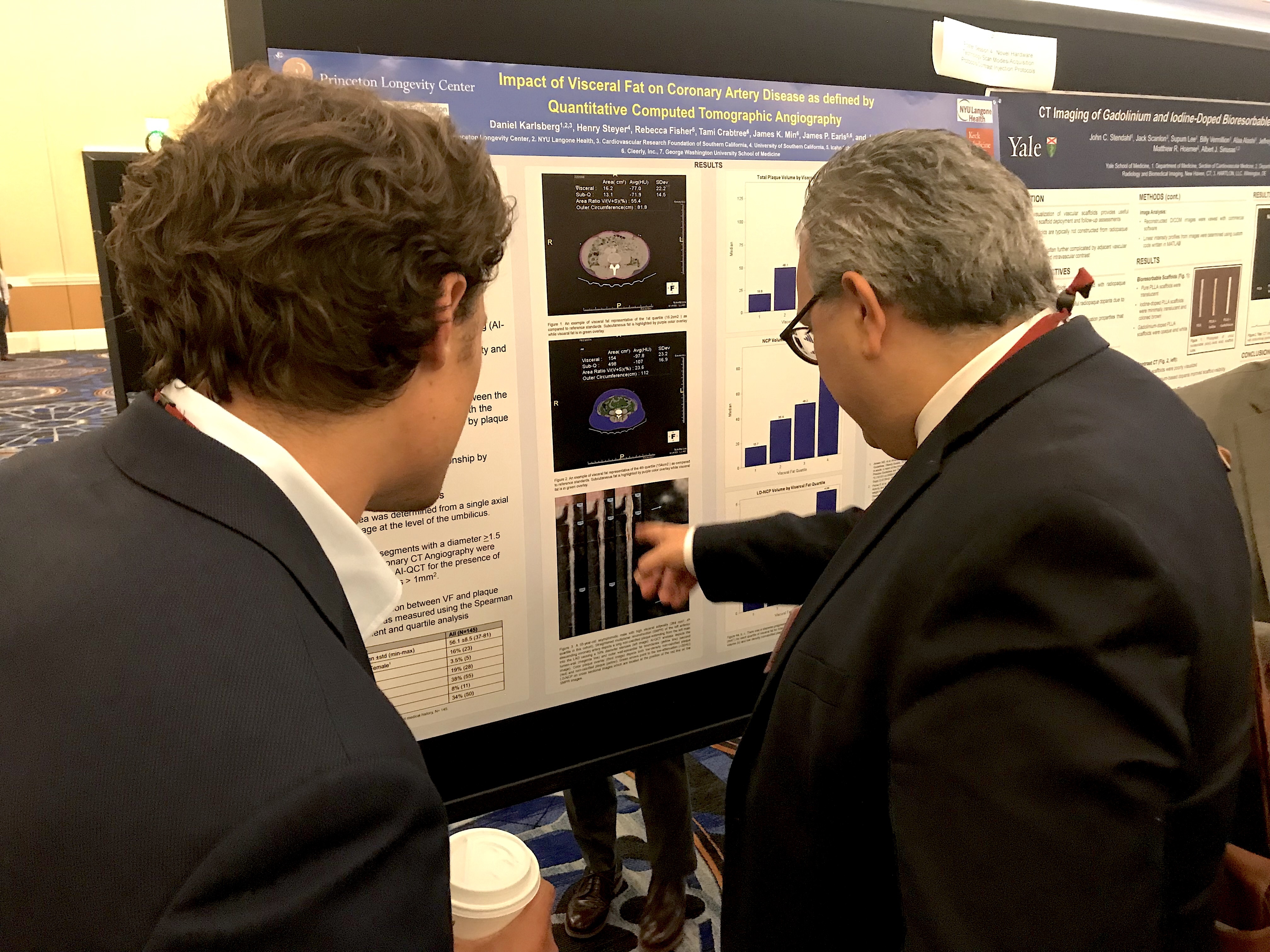
Poster presentation on the expo floor at SCCT.
Spectral CT that is built into photon-counting CT allows calcium to be removed or dialed down from coronary vessels so the plaque within the lumen can be viewed without calcium blooming. You can see the lumen in two heavily calcified coronary artery segments in the image demonstrated by Siemens healthcare at SCCT. Read and see more on this technology
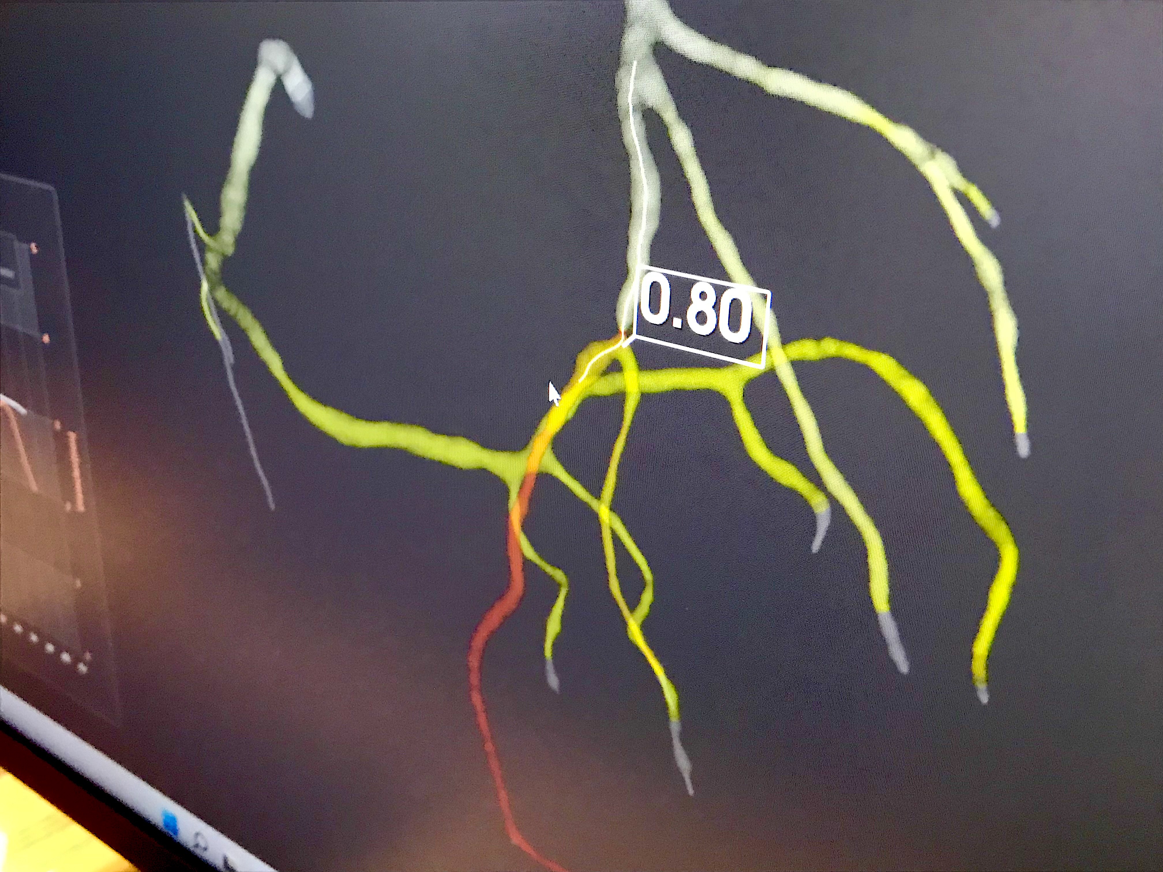
South Korean start-up artificial intelligence company AIMedic displayed its work-in-progress CT angiography algorithm that can derive FFR-CT values for the entire coronary tree in about 30 minutes. It uses an on-site server at the hospital or clinic, rather that requiring exams to be sent to third-parties off site. The company does not have FDA and was gathering feedback from physicians at SCCT.
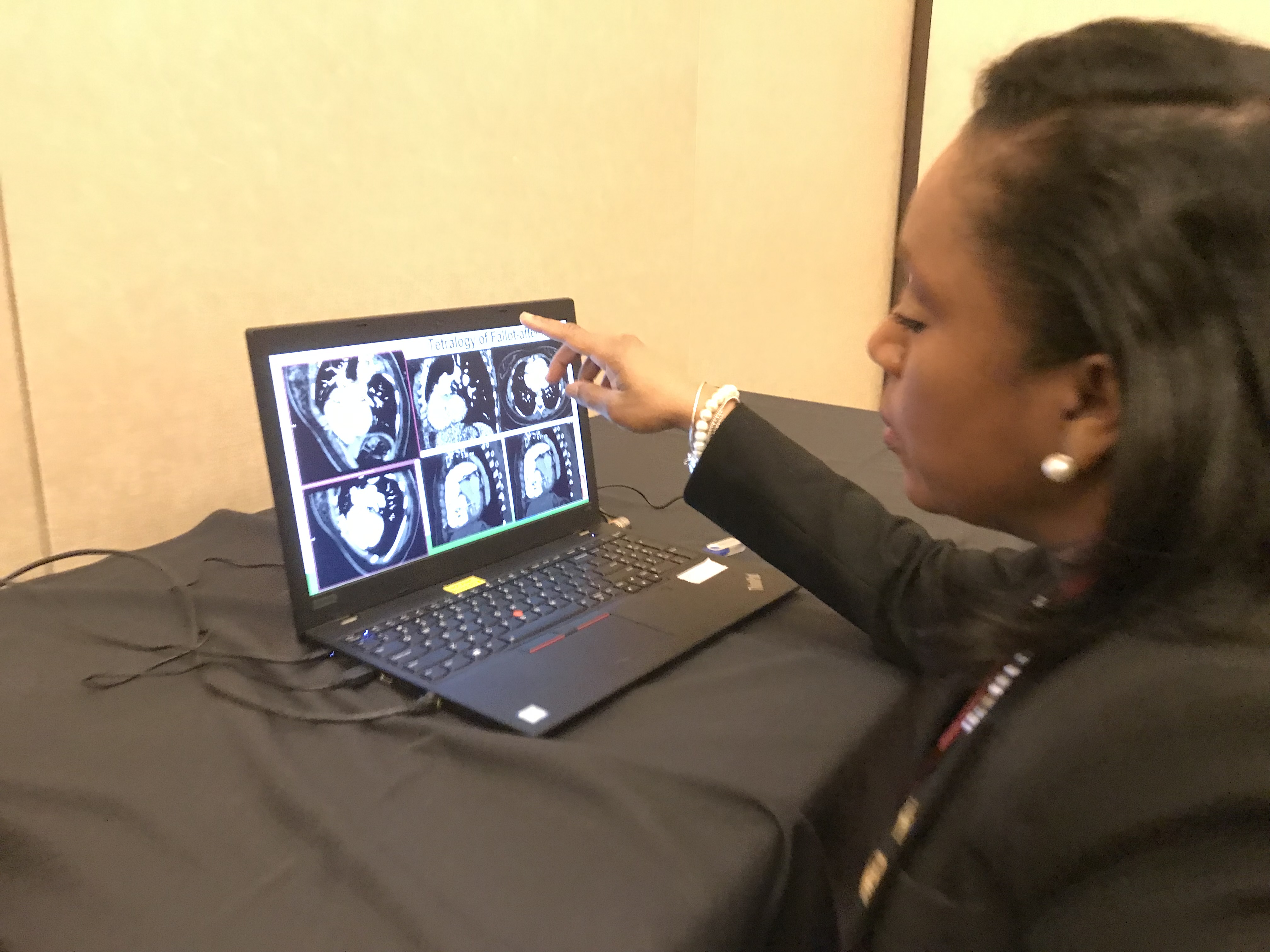
Renee Bullock-Palmer, MD, FACC, FAHA, FASNC, FASE, FSCCT, director of noninvasive cardiac imaging and the women's center at Deborah Heart and Lung Center, explains a complex congenital case. She presented on how CTA can be used to help assess congenital patients and better understand the anatomy prior to interventions.
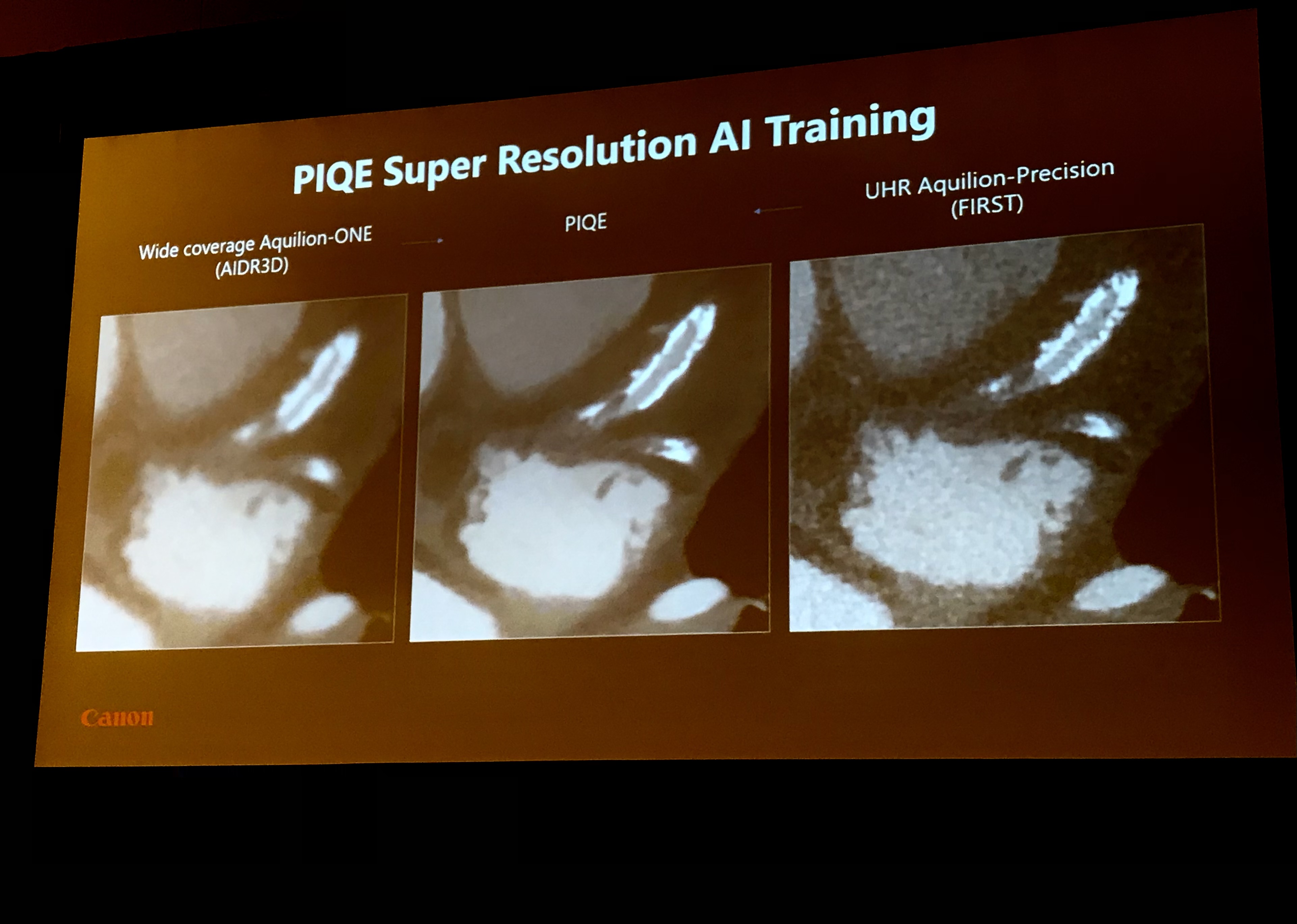
Example showing the difference in image resolution using artificial intelligence (AI) for image reconstruction compared to conventional iterative reconstruction. This example shows Canon's AI-based PIQE CT image reconstruction technology shown in a CT new innovations session. All the major CT vendors are developing AI image reconstruction to greatly improve resolution while enabling lower does CT scans.
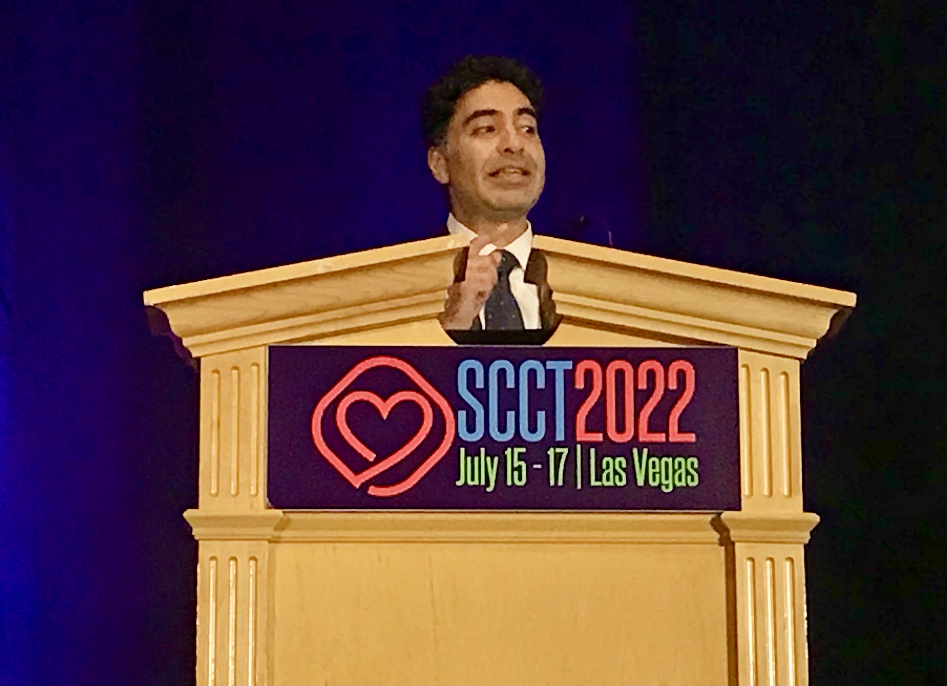
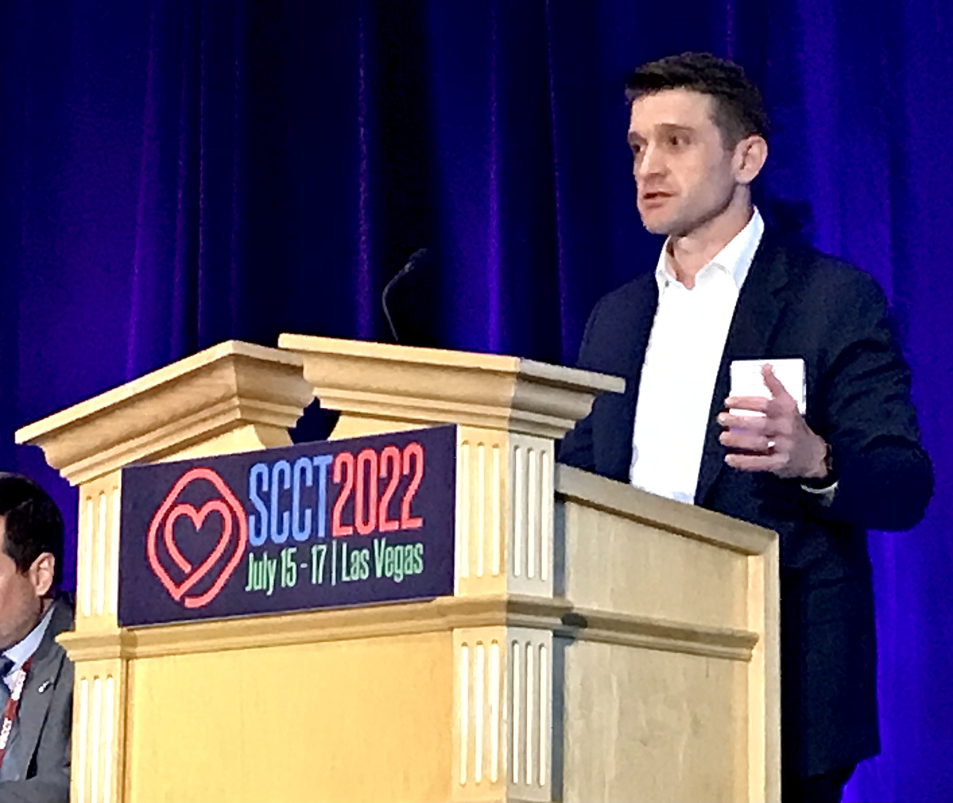
Incoming SCCT President and this year's meeting chairman Brian Ghoshharja, MD, Mass General, explains his hopes for the society in the coming year. Hear him explain why CTA has seen a big boost recently in the VIDEO: The new role of cardiac CT in chest pain evaluation

Downstairs from SCCT in the Caesar's Palace casino.
Poster sessions on the expo floor at SCCT 2022.
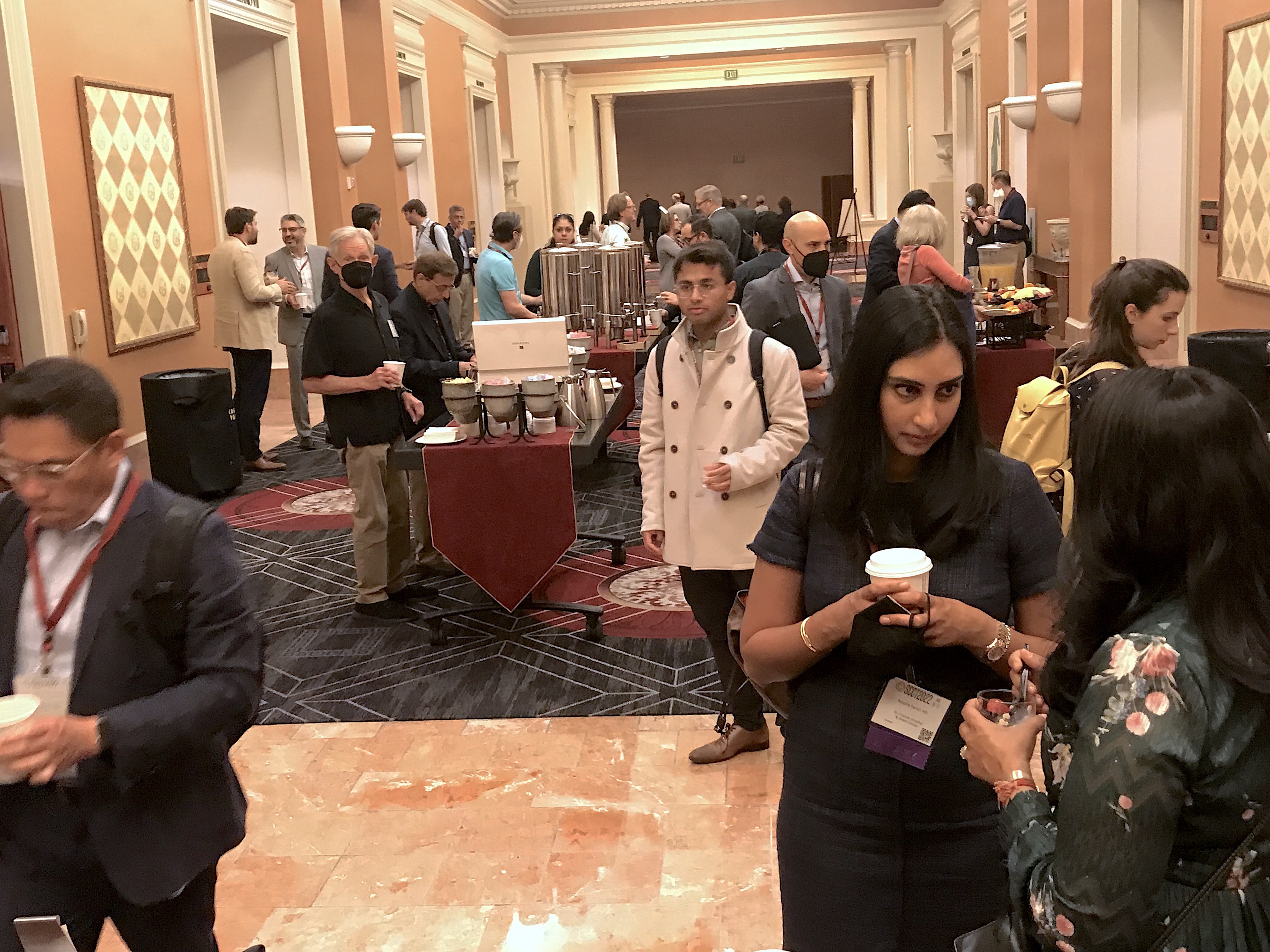
Attendees socializing outside a breakfast session. It was the first in-person meeting for SCCT since 2019 due to the COVID pandemic. Attendees and leadership both said the in-person networking was missed in 2020 and 2021.
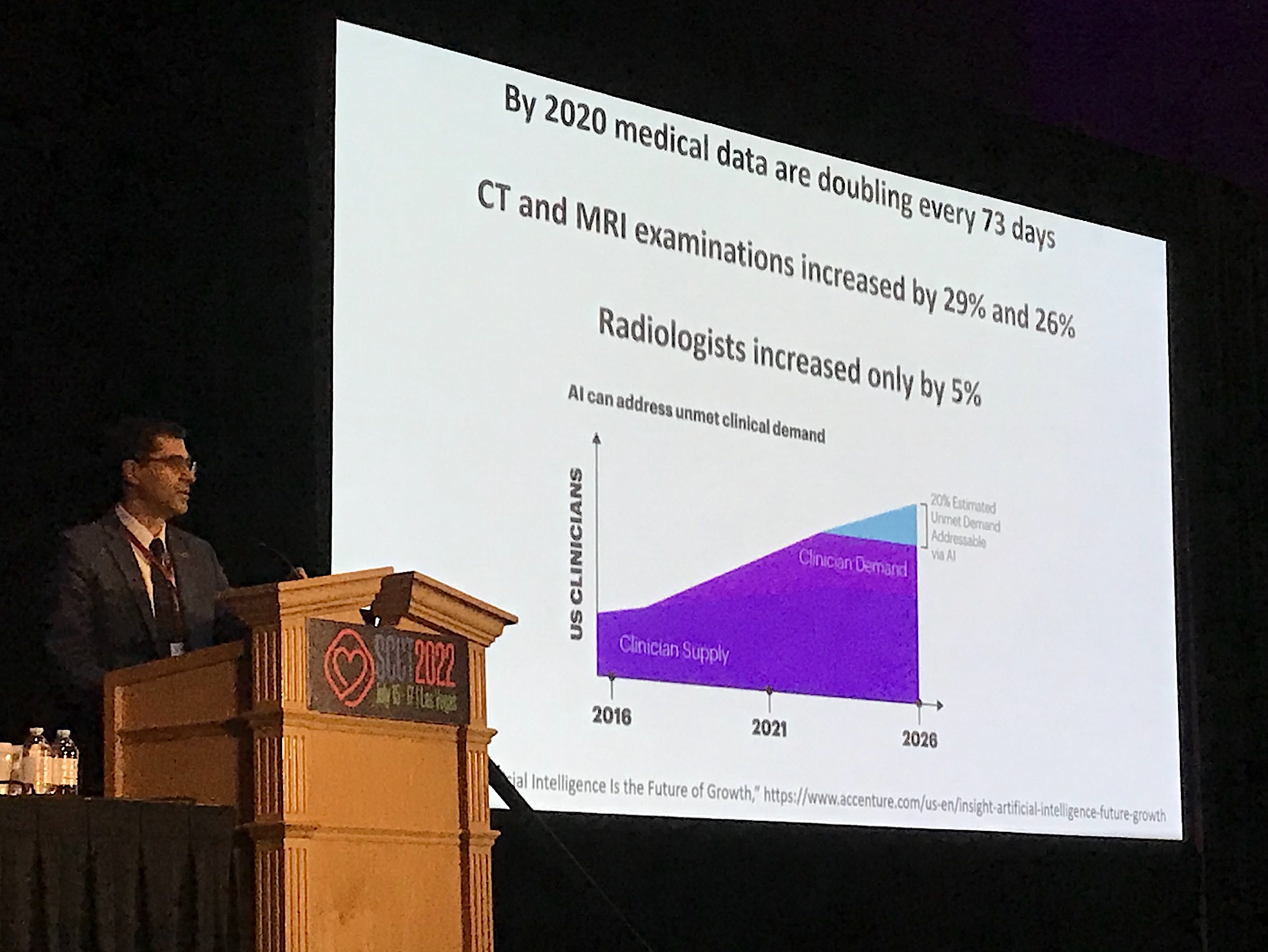
Carlo De Cecco, MD, PhD, FSCCT, associate professor of radiology and biomedical informatics at Emory University, explains considerations with artificial intelligence and why it is needed to help overcome growing radiologist and cardiologist shortages and where AI will fit in.

The iconic High Roller Ferris wheel and the monorail track in Las Vegas.
VA-ECMO can cause issues in cardiac CT evaluations because it changes the blood flow patterns in the body. This example was presented in a sesssion by Jody Shen, MD, Stanford. They show how contrast does not flow normally and there can appear to be an obstruction like a clot, but it is caused by the reversal of flow by ECMO. She said to get the contrast to go lower in the body, you may have to reduce the ECMO flow during the exam.
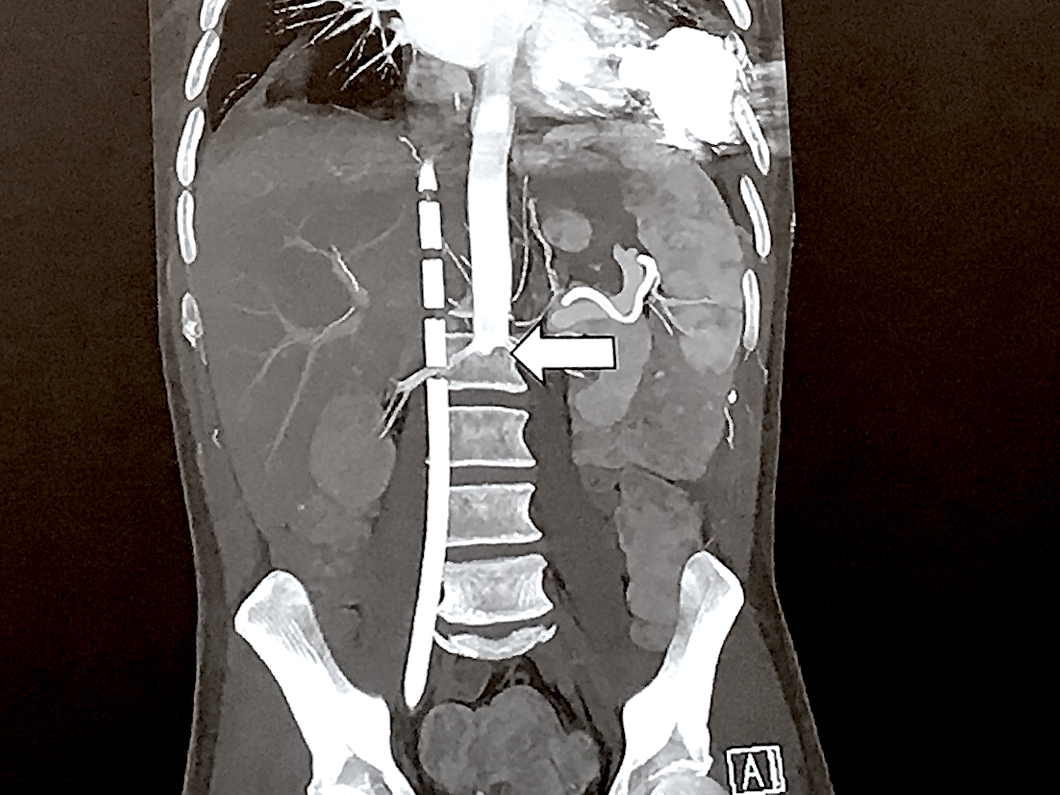
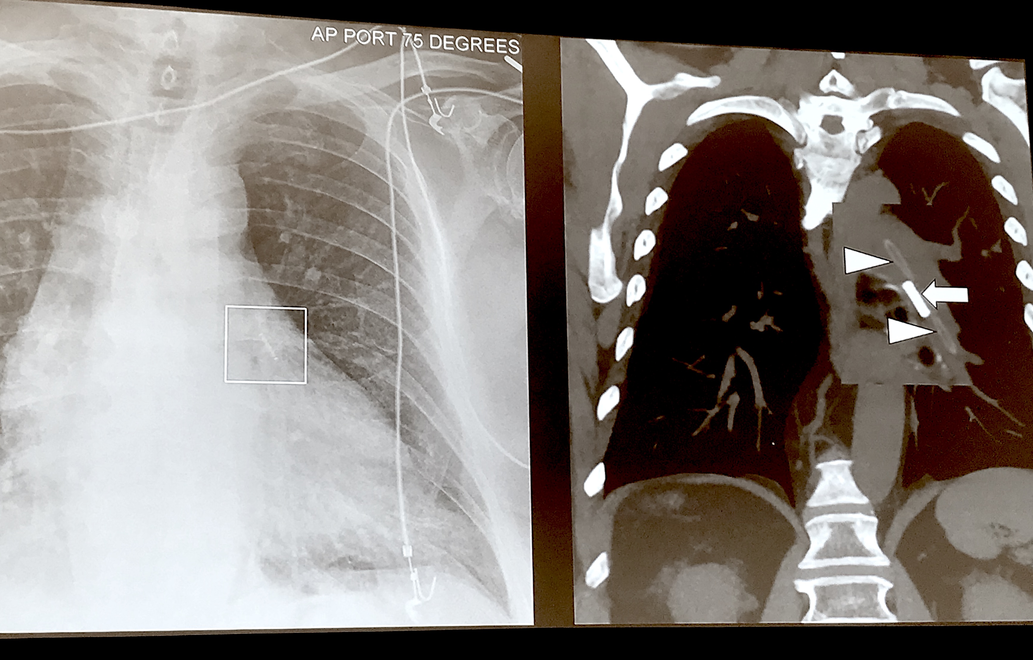
Example on CT of an intra-aortic balloon pump (IABP) accidentally positioned in the wrong vessel, resulting in aneurisms and pseudo aneurisms due to the excess pressure. Jody Shen, MD, Stanford, presented on uncommon device technologies that pop up on cardiac and CT exams that imagers need to be aware of.
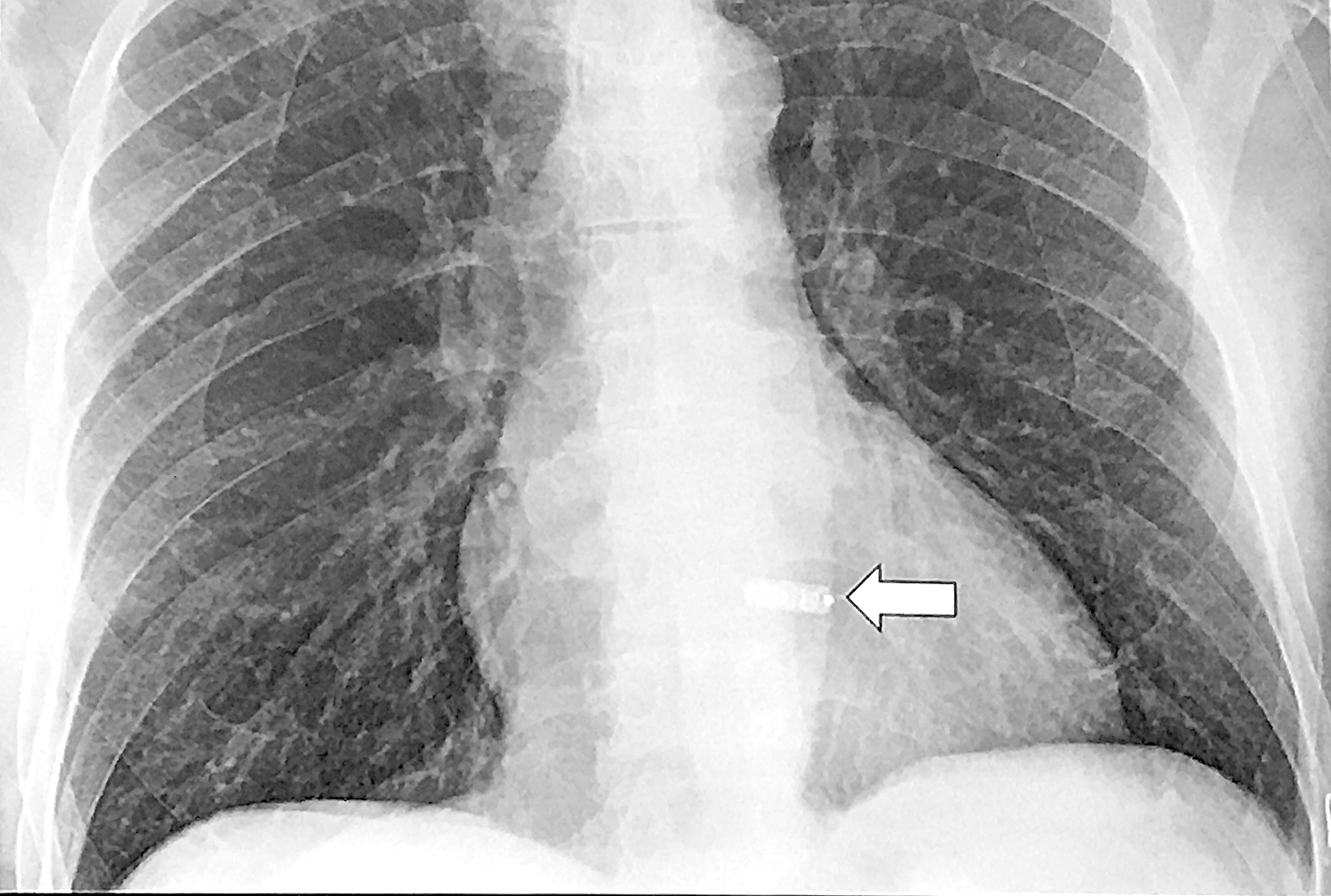
CT showing an intra-aortic balloon pump (IABP) inflated in the aortic arch. Jody Shen, MD, Stanford, presented on uncommon device technologies that pop up on cardiac and CT exams that imagers need to be aware of.
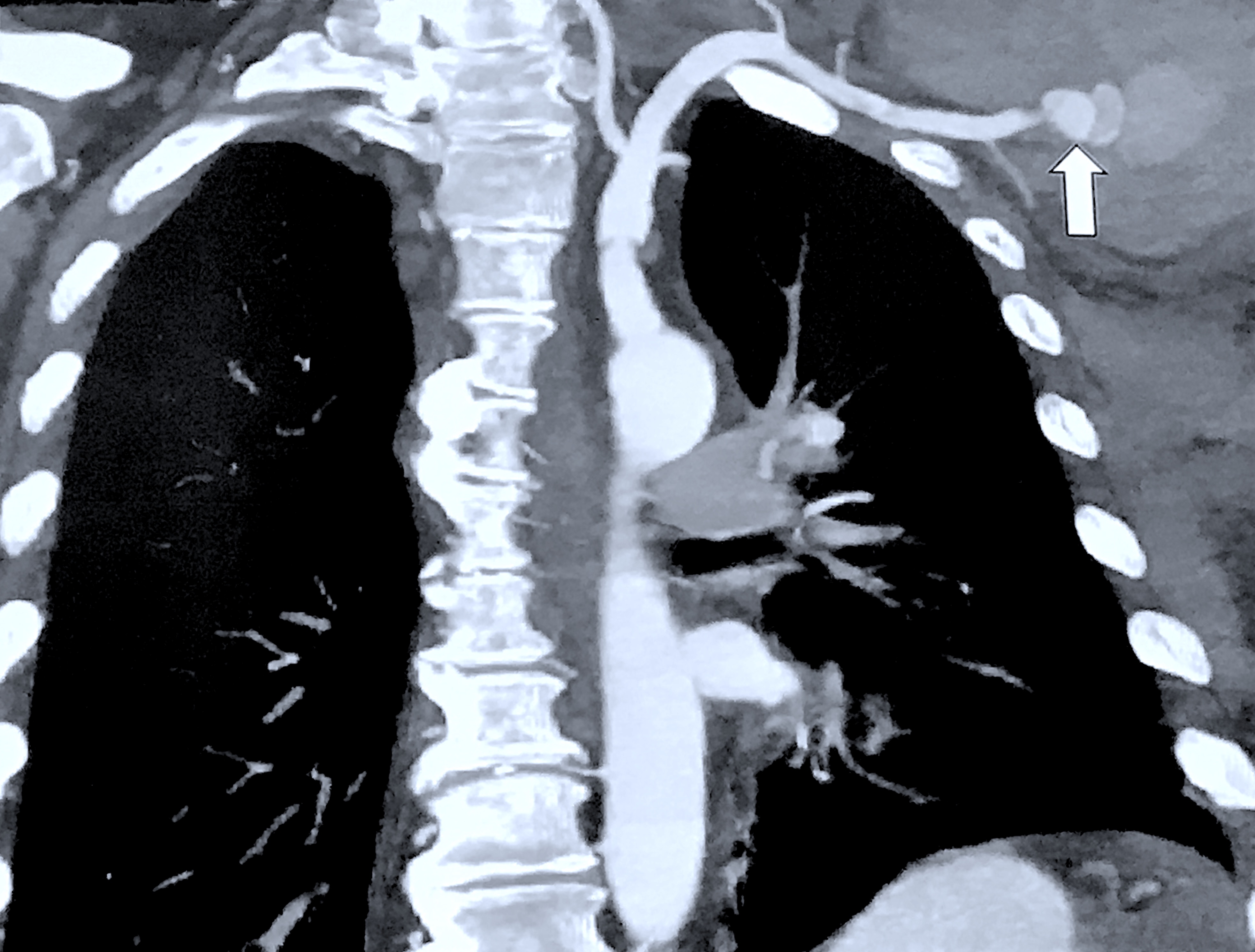
An example of a CardioMEMS implanted heart failure monitor in the pulmonary artery seen in a cardiac CT scan during a presentation at SCCT. Jody Shen, MD, Stanford, presented on uncommon device technologies that pop up on cardiac and CT exams that imagers need to be aware of. She said this device is identified by its wire loops attached at either end.
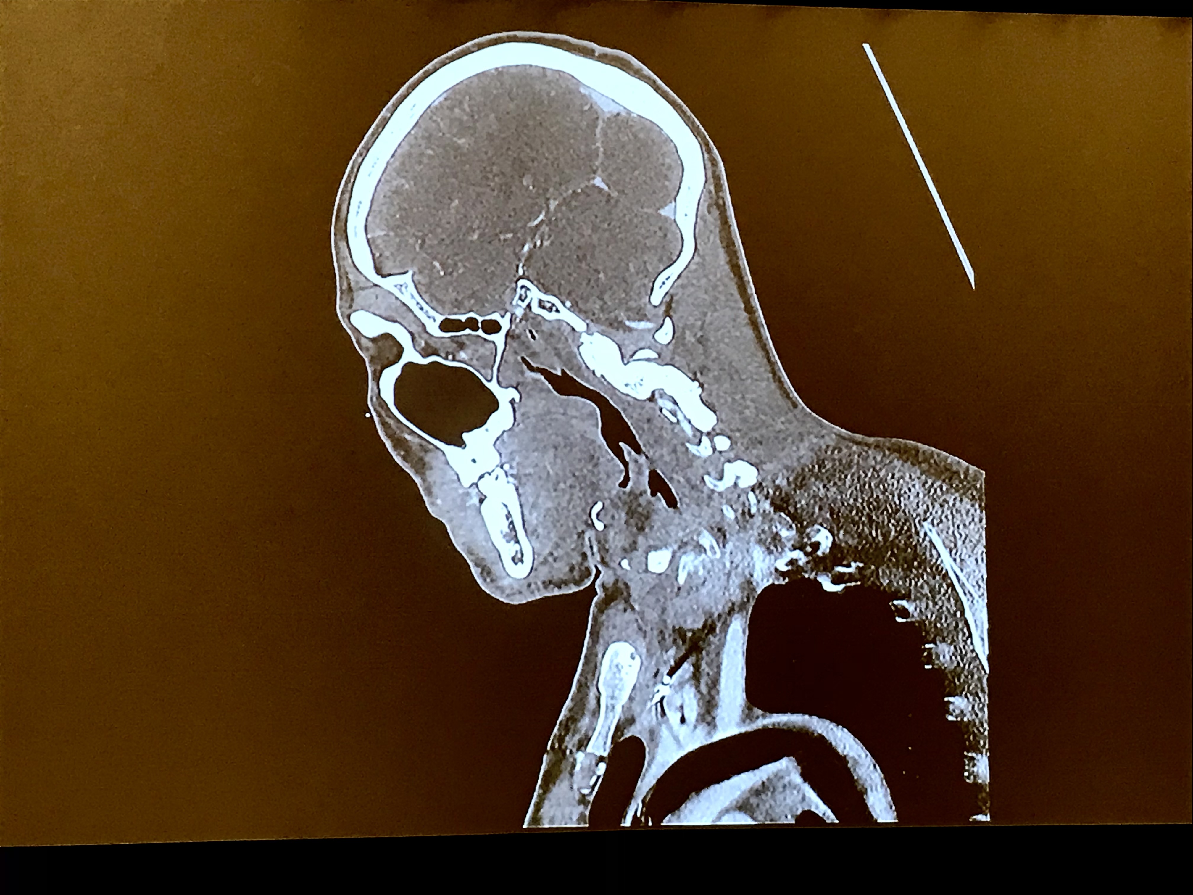
An example of a CT scout scan showing a metallic object inside the heart. Jody Shen, MD, Stanford, explained in her session this a transcatheter, leadless pacemaker, which will likely be a more common sight as these devices see wide use. She presented on uncommon device technologies that pop up on cardiac and CT exams that imagers need to be aware of.

Caesar's Palace Forum Shops area near where SCCT was hosted.

Caesar's Palace Forum Shops area near where SCCT was hosted.
Brent Savoie, MD, JD, vice chair for radiology informatics, section chief of cardiovascular imaging, Vanderbilt University, presents "Who will get sued when there is a misdiagnosis due to artificial intelligence?" session at SCCT. “The answer is everyone will get sued,” he explained. He outlined that the physician could share responsibility when AI fails, since they are ultimately responsible for diagnosis and reporting.
Attendees laugh at the act put on by leading CCTA experts who challenged each other to how fast they could read a CTA exam during the "Team Spirit" session. Most could pick out the key details within 10 seconds.
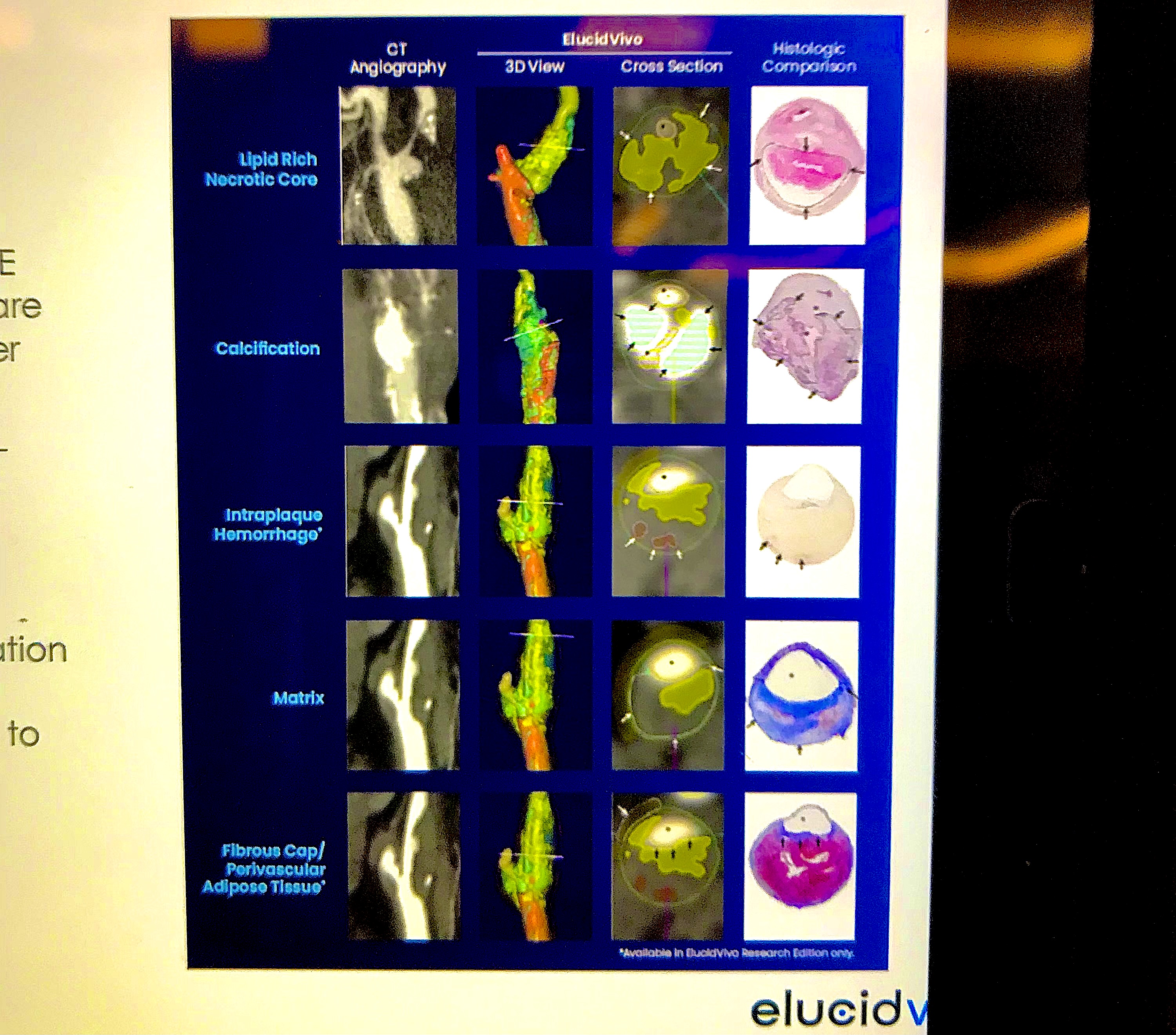
Cardiac artificial intelligence vendor Elucid has developed an algorithm to automatically assess a coronary CT scan to identify all the plaque in the vessels, classify it, and create a report with images. This is a clear technology direction with several vendors at the meeting this year, and it is embraced by the top experts here as the way of the future. They said this technology will allow serial CT assessment of patients to set a baseline and then track progression or regression of plaque based on drug therapies or interventions.
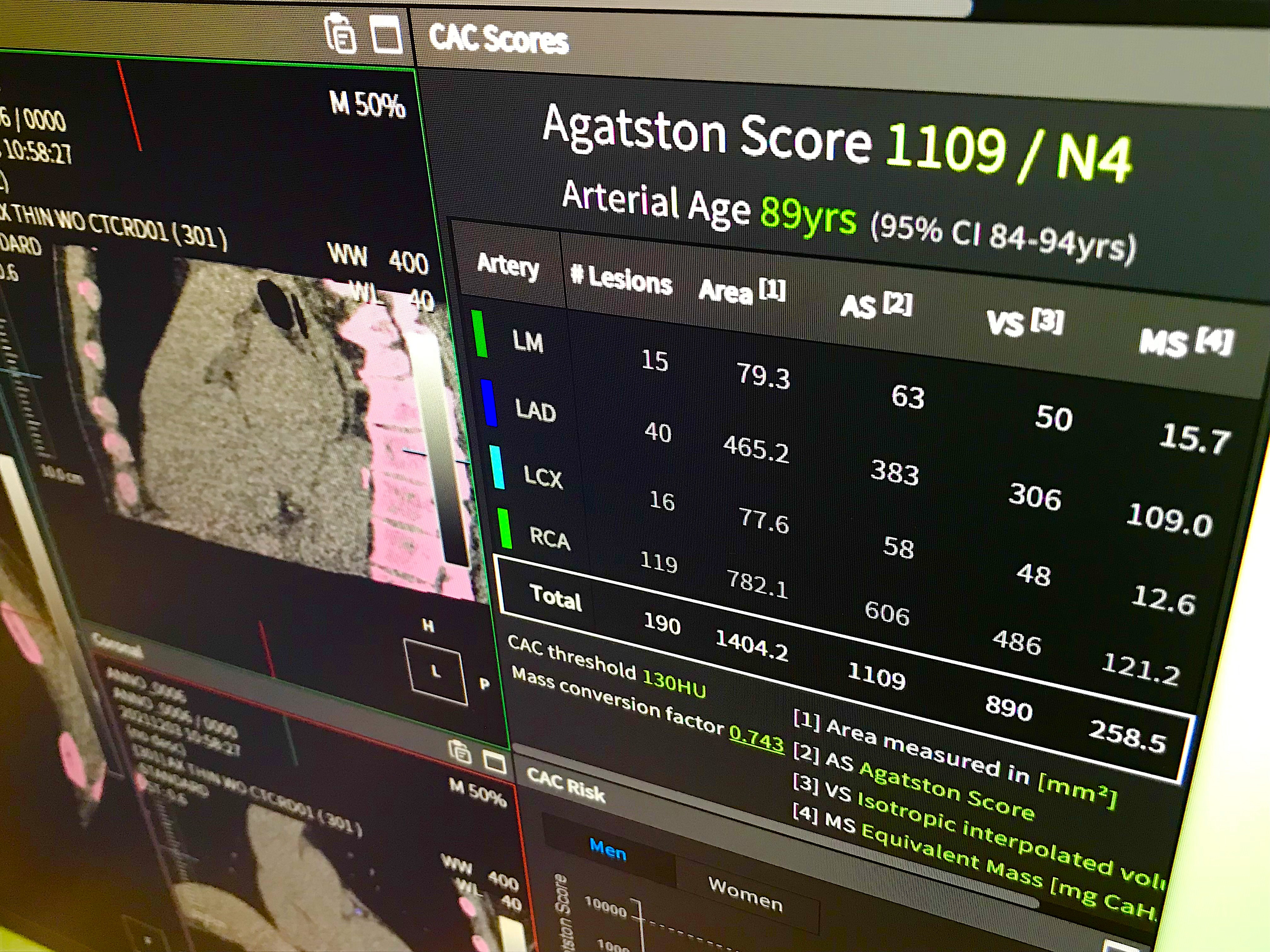
Example of an artificial intelligence (AI) fully automated report and quantitative review of a CT calcium scoring exam shown by the vendor Coreline at SCCT. CAC, or Agatston scoring, can offer patients a 10-year risk assessment for cardiac events. Some cardiologists use it as a way to convince patients to take statins, telling them if they have no calcium they can avoid stains for a few more years. But if they have calcium, the images and CAC score usually convince them the medication is a good idea. Many hospitals in the U.S. now offer CAC exams for $50-$150 if insurance does not cover it as a way to screen cardiac patients. The automation speeds the review and processing workflow, making them more appealing to radiology departments. Watch the VIDEO: Current guidelines for the use of CT calcium scoring in preventive cardiology
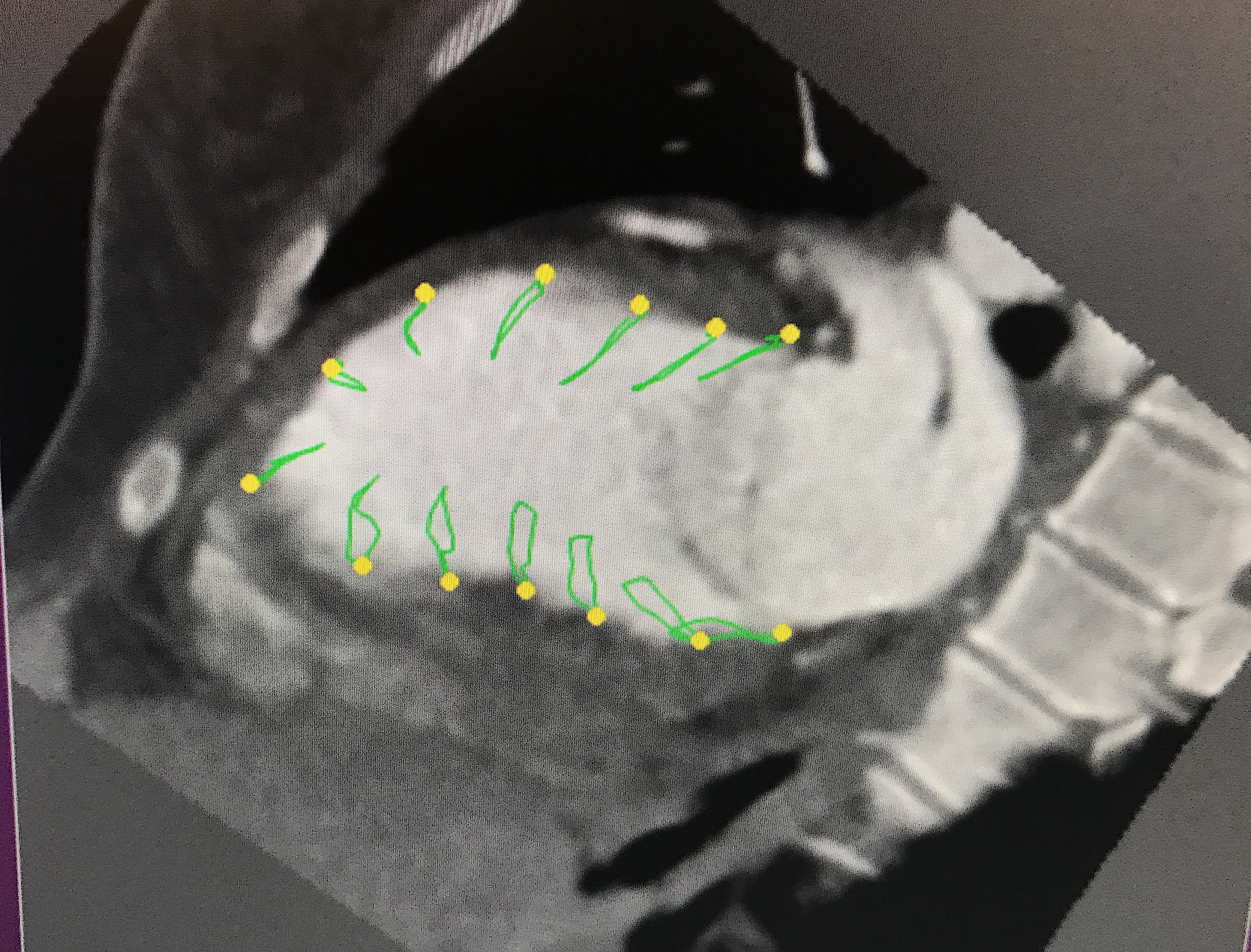
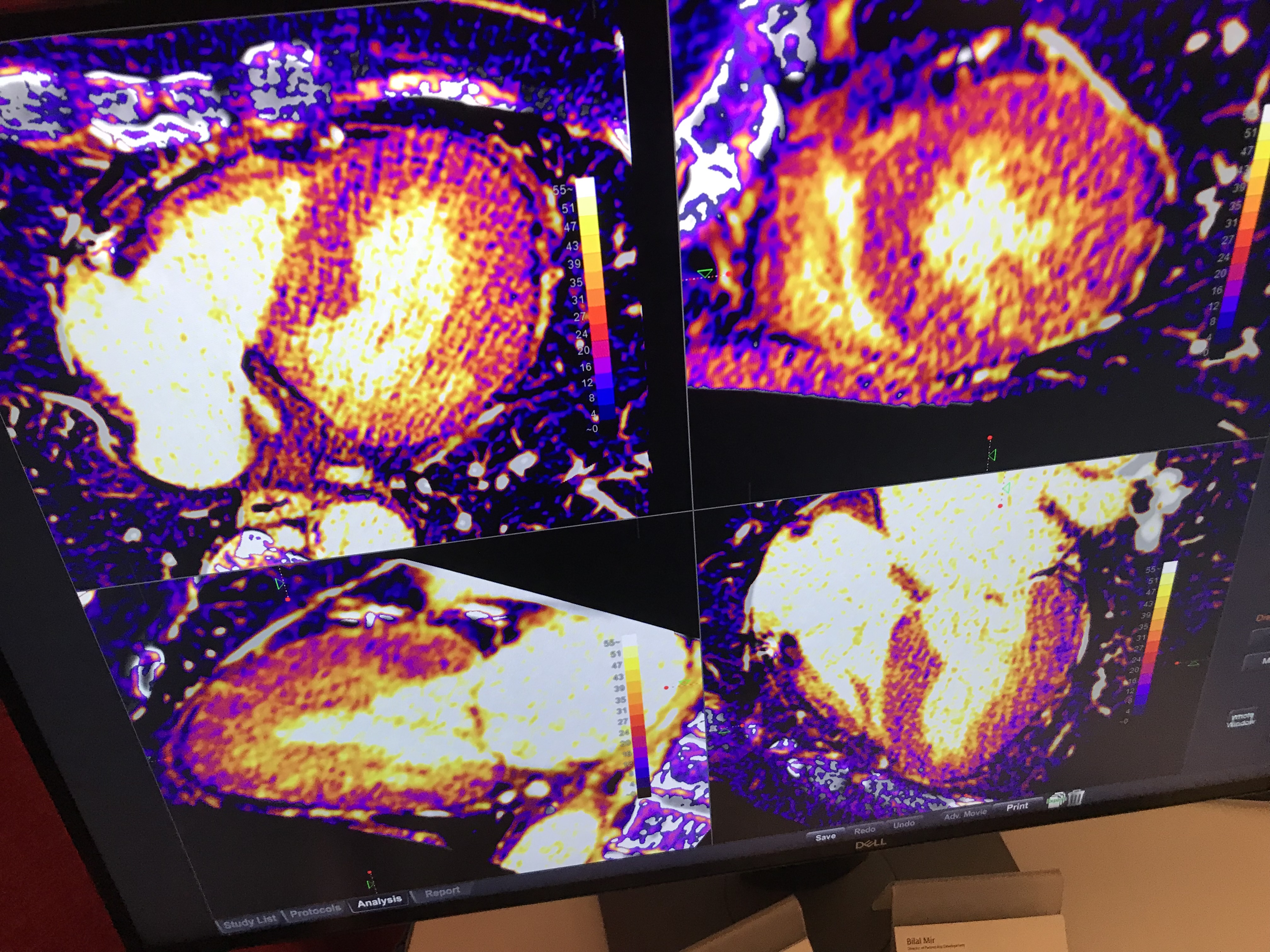
An example of cardiac CT myocardial strain imaging shown in sessions. The vendor Medis also demonstrated this technology of the show floor. It is similar to more commonly used echo strain imaging, which is used to gain more insight into a patient's cardiac function, and is a measure used to cardio-oncology monitoring of cardio-toxicity in patients under going cancer therapy. MRI is also commonly used in serial exam followup, but for patients who cannot undergo MRI, CT may offer a new option, said Joao Cavalcante, MD, Minneapolis Heart. He said about 40% of CABG patients also cannot get high quality echo exams needed for strain, so this technology allows them to be sent for a CT instead.

Attendees stop to catch up between sessions at SCCT.
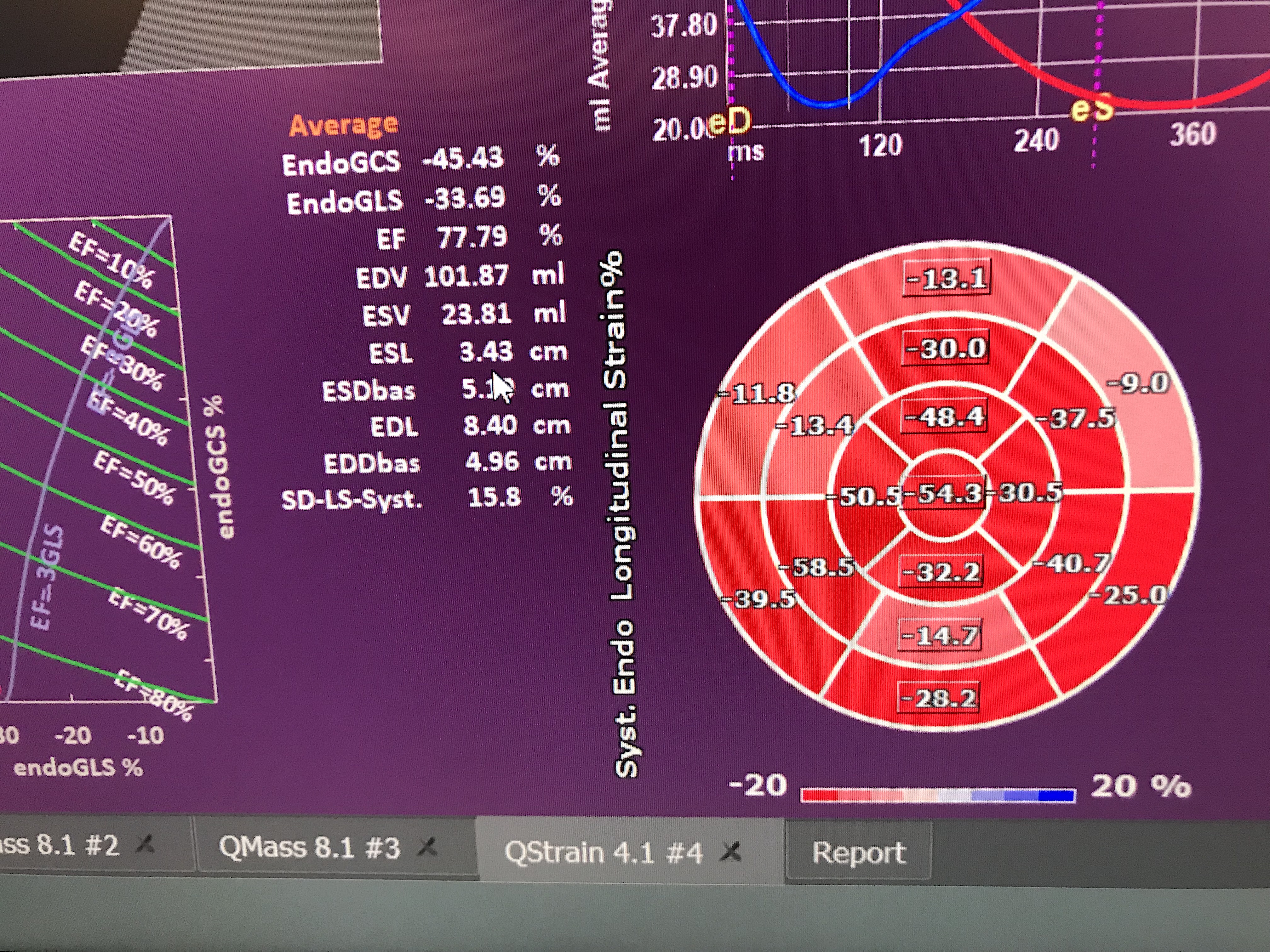
Bulls eye plot for a cardiac CT myocardial strain imaging exam from Medis.
Related Cardiac CT Imaging Content:
VIDEO: Cardiac CT now recommended as a front-line chest pain assessment tool
VIDEO: CT imaging to plan coronary interventions — Interview with Jon Leipsic
VIDEO: Office-based cardiac CT and FFR-CT offer a new business model
PHOTO GALLERY: Duly Health adopts outpatient cardiac CT as a standard of care
VIDEO: Use of CT to assess coronary plaques — Interview with Leslee Shaw, PhD
VIDEO: Top 6 takeaways from the Society of Cardiovascular CT 2022 meeting — interview with Eric Williamson, MD
VIDEO: The new role of cardiac CT under the 2021 chest pain evaluation guidelines — Interview with Eric Williamson, MD
VIDEO: Cardiac CT now recommended as a front-line chest pain assessment tool — Interview with Leslee Shaw, PhD
VIDEO: Office-based cardiac CT and FFR-CT offer a new business model
VIDEO: The new role of cardiac CT in chest pain evaluation — Interview with Brian Ghoshhajra, MD
CT a low-risk imaging alternative to invasive coronary angiography for suspected CAD
Photon-counting CT boosts image quality and reader confidence in identifying coronary artery disease
Radiologists launching new study assessing ‘one stop shop’ cardiac myocardial CT perfusion imaging

Dave Fornell has covered healthcare for more than 17 years, with a focus in cardiology and radiology. Fornell is a 5-time winner of a Jesse H. Neal Award, the most prestigious editorial honors in the field of specialized journalism. The wins included best technical content, best use of social media and best COVID-19 coverage. Fornell was also a three-time Neal finalist for best range of work by a single author. He produces more than 100 editorial videos each year, most of them interviews with key opinion leaders in medicine. He also writes technical articles, covers key trends, conducts video hospital site visits, and is very involved with social media. E-mail: [email protected]